4R1E

Crystal Structure of MTIP from Plasmodium falciparum in complex with a peptide-fragment chimera


Experimental Data Snapshot

  • Method: X-RAY DIFFRACTION
  • Resolution: 1.98 Å
  • R-Value Free: 0.223 
  • R-Value Work: 0.196 
  • R-Value Observed: 0.199 

wwPDB Validation   3D Report Full Report


Ligand Structure Quality Assessment 


This is version 1.4 of the entry. See complete history


Literature

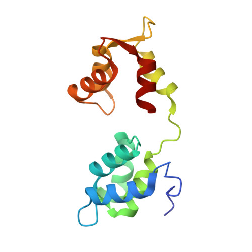
Targeting a Dynamic Protein-Protein Interaction: Fragment Screening against the Malaria Myosin A Motor Complex.

Douse, C.H.Vrielink, N.Wenlin, Z.Cota, E.Tate, E.W.

(2015) ChemMedChem 10: 134-143

  • DOI: https://doi.org/10.1002/cmdc.201402357
  • Primary Citation of Related Structures:  
    4R1E

  • PubMed Abstract: 

    Motility is a vital feature of the complex life cycle of Plasmodium falciparum, the apicomplexan parasite that causes human malaria. Processes such as host cell invasion are thought to be powered by a conserved actomyosin motor (containing myosin A or myoA), correct localization of which is dependent on a tight interaction with myosin A tail domain interacting protein (MTIP) at the inner membrane of the parasite. Although disruption of this protein-protein interaction represents an attractive means to investigate the putative roles of myoA-based motility and to inhibit the parasitic life cycle, no small molecules have been identified that bind to MTIP. Furthermore, it has not been possible to obtain a crystal structure of the free protein, which is highly dynamic and unstable in the absence of its natural myoA tail partner. Herein we report the de novo identification of the first molecules that bind to and stabilize MTIP via a fragment-based, integrated biophysical approach and structural investigations to examine the binding modes of hit compounds. The challenges of targeting such a dynamic system with traditional fragment screening workflows are addressed throughout.


  • Organizational Affiliation

    Department of Chemistry, Imperial College London, South Kensington, London SW7 2AZ (UK); Centre for Structural Biology, Department of Life Sciences, Imperial College London, South Kensington, London SW7 2AZ (UK); Institute of Chemical Biology, Imperial College London, South Kensington, London SW7 2AZ (UK). christopher.douse@googlemail.com.


Macromolecules
Find similar proteins by:  (by identity cutoff)  |  3D Structure
Entity ID: 1
MoleculeChains Sequence LengthOrganismDetailsImage
Myosin A tail domain interacting protein145Plasmodium falciparum 3D7Mutation(s): 0 
Gene Names: MTIPPFL2225w
UniProt
Find proteins for Q8I4W8 (Plasmodium falciparum (isolate 3D7))
Explore Q8I4W8 
Go to UniProtKB:  Q8I4W8
Entity Groups  
Sequence Clusters30% Identity50% Identity70% Identity90% Identity95% Identity100% Identity
UniProt GroupQ8I4W8
Sequence Annotations
Expand
  • Reference Sequence

Find similar proteins by:  Sequence   |   3D Structure  

Entity ID: 2
MoleculeChains Sequence LengthOrganismDetailsImage
Myosin-A15Plasmodium falciparum 3D7Mutation(s): 0 
UniProt
Find proteins for Q8IDR3 (Plasmodium falciparum (isolate 3D7))
Explore Q8IDR3 
Go to UniProtKB:  Q8IDR3
Entity Groups  
Sequence Clusters30% Identity50% Identity70% Identity90% Identity95% Identity100% Identity
UniProt GroupQ8IDR3
Sequence Annotations
Expand
  • Reference Sequence
Small Molecules
Ligands 1 Unique
IDChains Name / Formula / InChI Key2D Diagram3D Interactions
3EC
Query on 3EC

Download Ideal Coordinates CCD File 
C [auth B]5-{[(2-aminoethyl)sulfanyl]methyl}furan-2-carbaldehyde
C8 H11 N O2 S
IXLKFYGLYDVEMS-UHFFFAOYSA-N
Experimental Data & Validation

Experimental Data

  • Method: X-RAY DIFFRACTION
  • Resolution: 1.98 Å
  • R-Value Free: 0.223 
  • R-Value Work: 0.196 
  • R-Value Observed: 0.199 
  • Space Group: P 21 21 21
Unit Cell:
Length ( Å )Angle ( ˚ )
a = 36.71α = 90
b = 55.76β = 90
c = 75.86γ = 90
Software Package:
Software NamePurpose
Adxvdata processing
PHASERphasing
REFMACrefinement
MOSFLMdata reduction
SCALAdata scaling

Structure Validation

View Full Validation Report


Ligand Structure Quality Assessment 


Entry History 

Deposition Data

Revision History  (Full details and data files)

  • Version 1.0: 2014-11-12
    Type: Initial release
  • Version 1.1: 2015-01-14
    Changes: Database references
  • Version 1.2: 2017-08-23
    Changes: Data collection
  • Version 1.3: 2017-11-22
    Changes: Refinement description
  • Version 1.4: 2023-09-20
    Changes: Data collection, Database references, Derived calculations, Refinement description