4QZS

Crystal structure of the first bromodomain of human 3-fluoro tyrosine-labeled brd4 in complex with jq1


Experimental Data Snapshot

  • Method: X-RAY DIFFRACTION
  • Resolution: 1.45 Å
  • R-Value Free: 0.185 
  • R-Value Work: 0.140 

wwPDB Validation   3D Report Full Report


Ligand Structure Quality Assessment 


This is version 1.2 of the entry. See complete history


Literature

Fluorinated aromatic amino acids are sensitive (19)f NMR probes for bromodomain-ligand interactions.

Mishra, N.K.Urick, A.K.Ember, S.W.Schonbrunn, E.Pomerantz, W.C.

(2014) ACS Chem Biol 9: 2755-2760

  • DOI: https://doi.org/10.1021/cb5007344
  • Primary Citation of Related Structures:  
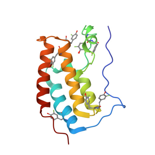
    4QZS

  • PubMed Abstract: 

    We describe a (19)F NMR method for detecting bromodomain-ligand interactions using fluorine-labeled aromatic amino acids due to the conservation of aromatic residues in the bromodomain binding site. We test the sensitivity, accuracy, and speed of this method with small molecule ligands (+)-JQ1, BI2536, Dinaciclib, TG101348, and acetaminophen using three bromodomains Brd4, BrdT, and BPTF. Simplified (19)F NMR spectra allowed for simultaneous testing of multiple bromodomains to assess selectivity and identification of a new BPTF ligand. Fluorine labeling only modestly affected the Brd4 structure and function assessed by isothermal titration calorimetry, circular dichroism, and X-ray crystallography. The speed, ease of interpretation, and low concentration of protein needed for binding experiments affords a new method to discover and characterize both native and new ligands.


  • Organizational Affiliation

    Department of Chemistry, University of Minnesota , 207 Pleasant St. SE, Minneapolis, Minnesota 55455, United States.


Macromolecules
Find similar proteins by:  (by identity cutoff)  |  3D Structure
Entity ID: 1
MoleculeChains Sequence LengthOrganismDetailsImage
Bromodomain-containing protein 4127Homo sapiensMutation(s): 0 
Gene Names: BRD4HUNK1
UniProt & NIH Common Fund Data Resources
Find proteins for O60885 (Homo sapiens)
Explore O60885 
Go to UniProtKB:  O60885
PHAROS:  O60885
GTEx:  ENSG00000141867 
Entity Groups  
Sequence Clusters30% Identity50% Identity70% Identity90% Identity95% Identity100% Identity
UniProt GroupO60885
Sequence Annotations
Expand
  • Reference Sequence
Small Molecules
Ligands 3 Unique
IDChains Name / Formula / InChI Key2D Diagram3D Interactions
JQ1
Query on JQ1

Download Ideal Coordinates CCD File 
B [auth A](6S)-6-(2-tert-butoxy-2-oxoethyl)-4-(4-chlorophenyl)-2,3,9-trimethyl-6,7-dihydrothieno[3,2-f][1,2,4]triazolo[4,3-a][1,4]diazepin-10-ium
C23 H26 Cl N4 O2 S
DNVXATUJJDPFDM-KRWDZBQOSA-O
EDO
Query on EDO

Download Ideal Coordinates CCD File 
C [auth A],
D [auth A]
1,2-ETHANEDIOL
C2 H6 O2
LYCAIKOWRPUZTN-UHFFFAOYSA-N
CL
Query on CL

Download Ideal Coordinates CCD File 
E [auth A]CHLORIDE ION
Cl
VEXZGXHMUGYJMC-UHFFFAOYSA-M
Modified Residues  1 Unique
IDChains TypeFormula2D DiagramParent
YOF
Query on YOF
A
L-PEPTIDE LINKINGC9 H10 F N O3TYR
Binding Affinity Annotations 
IDSourceBinding Affinity
JQ1 Binding MOAD:  4QZS Kd: 89 (nM) from 1 assay(s)
Experimental Data & Validation

Experimental Data

  • Method: X-RAY DIFFRACTION
  • Resolution: 1.45 Å
  • R-Value Free: 0.185 
  • R-Value Work: 0.140 
  • Space Group: P 21 21 21
Unit Cell:
Length ( Å )Angle ( ˚ )
a = 36.767α = 90
b = 44.898β = 90
c = 78.597γ = 90
Software Package:
Software NamePurpose
SERGUIdata collection
PHASERphasing
PHENIXrefinement
HKL-2000data reduction
HKL-2000data scaling

Structure Validation

View Full Validation Report


Ligand Structure Quality Assessment 


Entry History 

Deposition Data

Revision History  (Full details and data files)

  • Version 1.0: 2014-10-29
    Type: Initial release
  • Version 1.1: 2015-01-21
    Changes: Database references
  • Version 1.2: 2023-09-20
    Changes: Data collection, Database references, Derived calculations, Refinement description