4QTC

Structure of human haspin (GSG2) in complex with SCH772984 revealing the first type-I binding mode


Experimental Data Snapshot

  • Method: X-RAY DIFFRACTION
  • Resolution: 1.40 Å
  • R-Value Free: 0.164 
  • R-Value Work: 0.149 
  • R-Value Observed: 0.150 

wwPDB Validation   3D Report Full Report


Ligand Structure Quality Assessment 


This is version 1.2 of the entry. See complete history


Literature

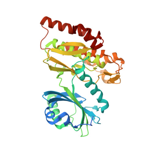
A unique inhibitor binding site in ERK1/2 is associated with slow binding kinetics.

Chaikuad, A.M C Tacconi, E.Zimmer, J.Liang, Y.Gray, N.S.Tarsounas, M.Knapp, S.

(2014) Nat Chem Biol 10: 853-860

  • DOI: https://doi.org/10.1038/nchembio.1629
  • Primary Citation of Related Structures:  
    4QTA, 4QTB, 4QTC, 4QTD, 4QTE

  • PubMed Abstract: 

    Activation of the ERK pathway is a hallmark of cancer, and targeting of upstream signaling partners led to the development of approved drugs. Recently, SCH772984 has been shown to be a selective and potent ERK1/2 inhibitor. Here we report the structural mechanism for its remarkable selectivity. In ERK1/2, SCH772984 induces a so-far-unknown binding pocket that accommodates the piperazine-phenyl-pyrimidine decoration. This new binding pocket was created by an inactive conformation of the phosphate-binding loop and an outward tilt of helix αC. In contrast, structure determination of SCH772984 with the off-target haspin and JNK1 revealed two canonical but distinct type I binding modes. Notably, the new binding mode with ERK1/2 was associated with slow binding kinetics in vitro as well as in cell-based assay systems. The described binding mode of SCH772984 with ERK1/2 enables the design of a new type of specific kinase inhibitors with prolonged on-target activity.


  • Organizational Affiliation

    Structural Genomics Consortium, University of Oxford, Old Road Campus Research Building, Oxford, UK.


Macromolecules
Find similar proteins by:  (by identity cutoff)  |  3D Structure
Entity ID: 1
MoleculeChains Sequence LengthOrganismDetailsImage
Serine/threonine-protein kinase haspin357Homo sapiensMutation(s): 0 
Gene Names: GSG2
EC: 2.7.11.1
UniProt & NIH Common Fund Data Resources
Find proteins for Q8TF76 (Homo sapiens)
Explore Q8TF76 
Go to UniProtKB:  Q8TF76
PHAROS:  Q8TF76
GTEx:  ENSG00000177602 
Entity Groups  
Sequence Clusters30% Identity50% Identity70% Identity90% Identity95% Identity100% Identity
UniProt GroupQ8TF76
Sequence Annotations
Expand
  • Reference Sequence
Small Molecules
Ligands 3 Unique
IDChains Name / Formula / InChI Key2D Diagram3D Interactions
38Z
Query on 38Z

Download Ideal Coordinates CCD File 
E [auth A](3R)-1-(2-oxo-2-{4-[4-(pyrimidin-2-yl)phenyl]piperazin-1-yl}ethyl)-N-[3-(pyridin-4-yl)-2H-indazol-5-yl]pyrrolidine-3-carboxamide
C33 H33 N9 O2
HDAJDNHIBCDLQF-RUZDIDTESA-N
MPD
Query on MPD

Download Ideal Coordinates CCD File 
B [auth A](4S)-2-METHYL-2,4-PENTANEDIOL
C6 H14 O2
SVTBMSDMJJWYQN-YFKPBYRVSA-N
GOL
Query on GOL

Download Ideal Coordinates CCD File 
C [auth A],
D [auth A]
GLYCEROL
C3 H8 O3
PEDCQBHIVMGVHV-UHFFFAOYSA-N
Binding Affinity Annotations 
IDSourceBinding Affinity
38Z Binding MOAD:  4QTC Kd: 297 (nM) from 1 assay(s)
Experimental Data & Validation

Experimental Data

  • Method: X-RAY DIFFRACTION
  • Resolution: 1.40 Å
  • R-Value Free: 0.164 
  • R-Value Work: 0.149 
  • R-Value Observed: 0.150 
  • Space Group: P 21 21 21
Unit Cell:
Length ( Å )Angle ( ˚ )
a = 77.89α = 90
b = 78.36β = 90
c = 81.75γ = 90
Software Package:
Software NamePurpose
GDAdata collection
PHASERphasing
REFMACrefinement
MOSFLMdata reduction
SCALAdata scaling

Structure Validation

View Full Validation Report


Ligand Structure Quality Assessment 


Entry History 

Deposition Data

Revision History  (Full details and data files)

  • Version 1.0: 2014-07-23
    Type: Initial release
  • Version 1.1: 2014-09-24
    Changes: Database references
  • Version 1.2: 2023-09-20
    Changes: Data collection, Database references, Derived calculations, Refinement description