4QT8

Crystal Structure of RON Sema-PSI-IPT1 extracellular domains in complex with MSP beta-chain


Experimental Data Snapshot

  • Method: X-RAY DIFFRACTION
  • Resolution: 3.00 Å
  • R-Value Free: 0.291 
  • R-Value Work: 0.234 
  • R-Value Observed: 0.237 

wwPDB Validation   3D Report Full Report


This is version 2.1 of the entry. See complete history


Literature

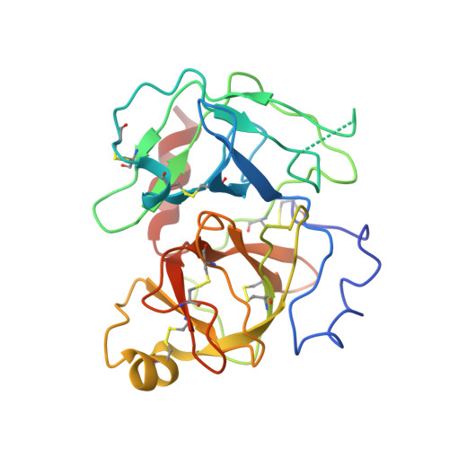
Structural basis for the binding specificity of human Recepteur d'Origine Nantais (RON) receptor tyrosine kinase to macrophage-stimulating protein.

Chao, K.L.Gorlatova, N.V.Eisenstein, E.Herzberg, O.

(2014) J Biol Chem 289: 29948-29960

  • DOI: https://doi.org/10.1074/jbc.M114.594341
  • Primary Citation of Related Structures:  
    4QT8

  • PubMed Abstract: 

    Recepteur d'origine nantais (RON) receptor tyrosine kinase and its ligand, serum macrophage-stimulating protein (MSP), play important roles in inflammation, cell growth, migration, and epithelial to mesenchymal transition during tumor development. The binding of mature MSPαβ (disulfide-linked α- and β-chains) to RON ectodomain modulates receptor dimerization, followed by autophosphorylation of tyrosines in the cytoplasmic receptor kinase domains. Receptor recognition is mediated by binding of MSP β-chain (MSPβ) to the RON Sema. Here we report the structure of RON Sema-PSI-IPT1 (SPI1) domains in complex with MSPβ at 3.0 Å resolution. The MSPβ serine protease-like β-barrel uses the degenerate serine protease active site to recognize blades 2, 3, and 4 of the β-propeller fold of RON Sema. Despite the sequence homology between RON and MET receptor tyrosine kinase and between MSP and hepatocyte growth factor, it is well established that there is no cross-reactivity between the two receptor-ligand systems. Comparison of the structure of RON SPI1 in complex with MSPβ and that of MET receptor tyrosine kinase Sema-PSI in complex with hepatocyte growth factor β-chain reveals the receptor-ligand selectivity determinants. Analytical ultracentrifugation studies of the SPI1-MSPβ interaction confirm the formation of a 1:1 complex. SPI1 and MSPαβ also associate primarily as a 1:1 complex with a binding affinity similar to that of SPI1-MSPβ. In addition, the SPI1-MSPαβ ultracentrifuge studies reveal a low abundance 2:2 complex with ∼ 10-fold lower binding affinity compared with the 1:1 species. These results support the hypothesis that the α-chain of MSPαβ mediates RON dimerization.


  • Organizational Affiliation

    From the Institute for Bioscience and Biotechnology Research, University of Maryland, Rockville, Maryland 20850 and.


Macromolecules
Find similar proteins by:  (by identity cutoff)  |  3D Structure
Entity ID: 1
MoleculeChains Sequence LengthOrganismDetailsImage
Macrophage-stimulating protein receptorA [auth B],
B [auth A]
669Homo sapiensMutation(s): 5 
Gene Names: MST1RPTK8RON
EC: 2.7.10.1
UniProt & NIH Common Fund Data Resources
Find proteins for Q04912 (Homo sapiens)
Explore Q04912 
Go to UniProtKB:  Q04912
PHAROS:  Q04912
GTEx:  ENSG00000164078 
Entity Groups  
Sequence Clusters30% Identity50% Identity70% Identity90% Identity95% Identity100% Identity
UniProt GroupQ04912
Sequence Annotations
Expand
  • Reference Sequence
Find similar proteins by:  (by identity cutoff)  |  3D Structure
Entity ID: 2
MoleculeChains Sequence LengthOrganismDetailsImage
Hepatocyte growth factor-like protein
C, D
253Homo sapiensMutation(s): 1 
Gene Names: D3F15S2DNF15S2HGFLMSI1MST1
UniProt & NIH Common Fund Data Resources
Find proteins for P26927 (Homo sapiens)
Explore P26927 
Go to UniProtKB:  P26927
PHAROS:  P26927
Entity Groups  
Sequence Clusters30% Identity50% Identity70% Identity90% Identity95% Identity100% Identity
UniProt GroupP26927
Sequence Annotations
Expand
  • Reference Sequence
Oligosaccharides

Help

Entity ID: 3
MoleculeChains Length2D Diagram Glycosylation3D Interactions
beta-D-mannopyranose-(1-4)-2-acetamido-2-deoxy-beta-D-glucopyranose-(1-4)-2-acetamido-2-deoxy-beta-D-glucopyranose
E
3N-Glycosylation
Glycosylation Resources
GlyTouCan:  G15407YE
GlyCosmos:  G15407YE
GlyGen:  G15407YE
Entity ID: 4
MoleculeChains Length2D Diagram Glycosylation3D Interactions
2-acetamido-2-deoxy-beta-D-glucopyranose-(1-4)-2-acetamido-2-deoxy-beta-D-glucopyranose
F
2N-Glycosylation
Glycosylation Resources
GlyTouCan:  G42666HT
GlyCosmos:  G42666HT
GlyGen:  G42666HT
Experimental Data & Validation

Experimental Data

  • Method: X-RAY DIFFRACTION
  • Resolution: 3.00 Å
  • R-Value Free: 0.291 
  • R-Value Work: 0.234 
  • R-Value Observed: 0.237 
  • Space Group: P 1 21 1
Unit Cell:
Length ( Å )Angle ( ˚ )
a = 106.585α = 90
b = 63.775β = 90.08
c = 146.049γ = 90
Software Package:
Software NamePurpose
Blu-Icedata collection
PHASERphasing
REFMACrefinement
XDSdata reduction
XSCALEdata scaling

Structure Validation

View Full Validation Report


Entry History 

Deposition Data

Revision History  (Full details and data files)

  • Version 1.0: 2014-09-17
    Type: Initial release
  • Version 1.1: 2014-09-24
    Changes: Database references
  • Version 1.2: 2015-04-01
    Changes: Database references
  • Version 1.3: 2017-11-22
    Changes: Refinement description
  • Version 2.0: 2020-07-29
    Type: Remediation
    Reason: Carbohydrate remediation
    Changes: Atomic model, Data collection, Database references, Derived calculations, Structure summary
  • Version 2.1: 2023-09-20
    Changes: Data collection, Database references, Refinement description, Structure summary