4QSH

Crystal Structure of L. monocytogenes Pyruvate Carboxylase in complex with Cyclic-di-AMP


Experimental Data Snapshot

  • Method: X-RAY DIFFRACTION
  • Resolution: 2.51 Å
  • R-Value Free: 0.233 
  • R-Value Work: 0.195 
  • R-Value Observed: 0.197 

wwPDB Validation   3D Report Full Report


Ligand Structure Quality Assessment 


This is version 1.1 of the entry. See complete history


Literature

The Cyclic Dinucleotide c-di-AMP Is an Allosteric Regulator of Metabolic Enzyme Function.

Sureka, K.Choi, P.H.Precit, M.Delince, M.Pensinger, D.A.Huynh, T.N.Jurado, A.R.Goo, Y.A.Sadilek, M.Iavarone, A.T.Sauer, J.D.Tong, L.Woodward, J.J.

(2014) Cell 158: 1389-1401

  • DOI: https://doi.org/10.1016/j.cell.2014.07.046
  • Primary Citation of Related Structures:  
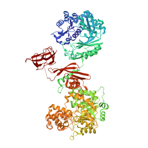
    4QSH, 4QSK, 4QSL

  • PubMed Abstract: 

    Cyclic di-adenosine monophosphate (c-di-AMP) is a broadly conserved second messenger required for bacterial growth and infection. However, the molecular mechanisms of c-di-AMP signaling are still poorly understood. Using a chemical proteomics screen for c-di-AMP-interacting proteins in the pathogen Listeria monocytogenes, we identified several broadly conserved protein receptors, including the central metabolic enzyme pyruvate carboxylase (LmPC). Biochemical and crystallographic studies of the LmPC-c-di-AMP interaction revealed a previously unrecognized allosteric regulatory site 25 Å from the active site. Mutations in this site disrupted c-di-AMP binding and affected catalytic activity of LmPC as well as PC from pathogenic Enterococcus faecalis. C-di-AMP depletion resulted in altered metabolic activity in L. monocytogenes. Correction of this metabolic imbalance rescued bacterial growth, reduced bacterial lysis, and resulted in enhanced bacterial burdens during infection. These findings greatly expand the c-di-AMP signaling repertoire and reveal a central metabolic regulatory role for a cyclic dinucleotide.


  • Organizational Affiliation

    Department of Microbiology, University of Washington, Seattle, WA 98195, USA.


Macromolecules
Find similar proteins by:  (by identity cutoff)  |  3D Structure
Entity ID: 1
MoleculeChains Sequence LengthOrganismDetailsImage
Pyruvate carboxylase
A, B, C, D
1,148Listeria monocytogenesMutation(s): 0 
Gene Names: AX24_02755
EC: 6.4.1.1
Entity Groups  
Sequence Clusters30% Identity50% Identity70% Identity90% Identity95% Identity100% Identity
Sequence Annotations
Expand
  • Reference Sequence
Small Molecules
Ligands 3 Unique
IDChains Name / Formula / InChI Key2D Diagram3D Interactions
2BA
Query on 2BA

Download Ideal Coordinates CCD File 
G [auth A],
H [auth A],
K [auth C]
(2R,3R,3aS,5R,7aR,9R,10R,10aS,12R,14aR)-2,9-bis(6-amino-9H-purin-9-yl)octahydro-2H,7H-difuro[3,2-d:3',2'-j][1,3,7,9,2,8 ]tetraoxadiphosphacyclododecine-3,5,10,12-tetrol 5,12-dioxide
C20 H24 N10 O12 P2
PDXMFTWFFKBFIN-XPWFQUROSA-N
FLC
Query on FLC

Download Ideal Coordinates CCD File 
E [auth A],
I [auth B],
L [auth C],
N [auth D]
CITRATE ANION
C6 H5 O7
KRKNYBCHXYNGOX-UHFFFAOYSA-K
MN
Query on MN

Download Ideal Coordinates CCD File 
F [auth A],
J [auth B],
M [auth C],
O [auth D]
MANGANESE (II) ION
Mn
WAEMQWOKJMHJLA-UHFFFAOYSA-N
Experimental Data & Validation

Experimental Data

  • Method: X-RAY DIFFRACTION
  • Resolution: 2.51 Å
  • R-Value Free: 0.233 
  • R-Value Work: 0.195 
  • R-Value Observed: 0.197 
  • Space Group: P 1 21 1
Unit Cell:
Length ( Å )Angle ( ˚ )
a = 96.778α = 90
b = 153.296β = 101.58
c = 221.24γ = 90
Software Package:
Software NamePurpose
PHASERphasing
REFMACrefinement
DENZOdata reduction
SCALEPACKdata scaling

Structure Validation

View Full Validation Report


Ligand Structure Quality Assessment 


Entry History 

Deposition Data

Revision History  (Full details and data files)

  • Version 1.0: 2014-10-29
    Type: Initial release
  • Version 1.1: 2023-09-20
    Changes: Data collection, Database references, Derived calculations, Refinement description, Structure summary