4QRY

the ground state and the N intermediate of pharaonis halorhodopsin in complex with bromide ion


Experimental Data Snapshot

  • Method: X-RAY DIFFRACTION
  • Resolution: 2.20 Å
  • R-Value Free: 0.243 
  • R-Value Work: 0.214 
  • R-Value Observed: 0.214 

wwPDB Validation   3D Report Full Report


Ligand Structure Quality Assessment 


This is version 1.2 of the entry. See complete history


Literature

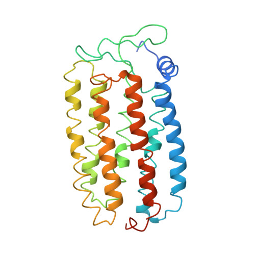
Crystal Structures of the L1, L2, N, and O States of pharaonis Halorhodopsin

Kouyama, T.Kawaguchi, H.Nakanishi, T.Kubo, H.Murakami, M.

(2015) Biophys J 108: 2680-2690

  • DOI: https://doi.org/10.1016/j.bpj.2015.04.027
  • Primary Citation of Related Structures:  
    4QRY

  • PubMed Abstract: 

    Halorhodopsin from Natronomonas pharaonis (pHR) functions as a light-driven halide ion pump. In the presence of halide ions, the photochemical reaction of pHR is described by the scheme: K→ L1 → L2 → N → O → pHR' → pHR. Here, we report light-induced structural changes of the pHR-bromide complex observed in the C2 crystal. In the L1-to-L2 transition, the bromide ion that initially exists in the extracellular vicinity of retinal moves across the retinal Schiff base. Upon the formation of the N state with a bromide ion bound to the cytoplasmic vicinity of the retinal Schiff base, the cytoplasmic half of helix F moves outward to create a water channel in the cytoplasmic interhelical space, whereas the extracellular half of helix C moves inward. During the transition from N to an N-like reaction state with retinal assuming the 13-cis/15-syn configuration, the translocated bromide ion is released into the cytoplasmic medium. Subsequently, helix F relaxes into its original conformation, generating the O state. Anion uptake from the extracellular side occurs when helix C relaxes into its original conformation. These structural data provide insight into the structural basis of unidirectional anion transport.


  • Organizational Affiliation

    Department of Physics, Graduate School of Science, Nagoya University, Nagoya, Japan; RIKEN Harima Branch, 1-1-1, Kouto, Sayo, Hyogo, Japan. Electronic address: kouyama@bio.phys.nagoya-u.ac.jp.


Macromolecules
Find similar proteins by:  (by identity cutoff)  |  3D Structure
Entity ID: 1
MoleculeChains Sequence LengthOrganismDetailsImage
Halorhodopsin291Natronomonas pharaonisMutation(s): 0 
Entity Groups  
Sequence Clusters30% Identity50% Identity70% Identity90% Identity95% Identity100% Identity
Sequence Annotations
Expand
  • Reference Sequence
Small Molecules
Ligands 4 Unique
IDChains Name / Formula / InChI Key2D Diagram3D Interactions
L2P
Query on L2P

Download Ideal Coordinates CCD File 
AA [auth F]
FA [auth G]
R [auth C]
V [auth E]
Y [auth F]
AA [auth F],
FA [auth G],
R [auth C],
V [auth E],
Y [auth F],
Z [auth F]
2,3-DI-PHYTANYL-GLYCEROL
C43 H88 O3
ISDBCJSGCHUHFI-UMZPFTBHSA-N
BNG
Query on BNG

Download Ideal Coordinates CCD File 
J [auth A],
K [auth B],
S [auth E],
T [auth E],
U [auth E]
nonyl beta-D-glucopyranoside
C15 H30 O6
QFAPUKLCALRPLH-UXXRCYHCSA-N
RET
Query on RET

Download Ideal Coordinates CCD File 
CA [auth F]
G [auth A]
GA [auth G]
L [auth B]
O [auth C]
CA [auth F],
G [auth A],
GA [auth G],
L [auth B],
O [auth C],
W [auth E]
RETINAL
C20 H28 O
NCYCYZXNIZJOKI-OVSJKPMPSA-N
BR
Query on BR

Download Ideal Coordinates CCD File 
BA [auth F]
DA [auth F]
EA [auth F]
H [auth A]
HA [auth G]
BA [auth F],
DA [auth F],
EA [auth F],
H [auth A],
HA [auth G],
I [auth A],
IA [auth G],
M [auth B],
N [auth B],
P [auth C],
Q [auth C],
X [auth E]
BROMIDE ION
Br
CPELXLSAUQHCOX-UHFFFAOYSA-M
Experimental Data & Validation

Experimental Data

  • Method: X-RAY DIFFRACTION
  • Resolution: 2.20 Å
  • R-Value Free: 0.243 
  • R-Value Work: 0.214 
  • R-Value Observed: 0.214 
  • Space Group: C 1 2 1
Unit Cell:
Length ( Å )Angle ( ˚ )
a = 153.36α = 90
b = 98.3β = 128.13
c = 100.53γ = 90
Software Package:
Software NamePurpose
ADSCdata collection
CNSrefinement
MOSFLMdata reduction
SCALAdata scaling
CNSphasing

Structure Validation

View Full Validation Report


Ligand Structure Quality Assessment 


Entry History 

Deposition Data

Revision History  (Full details and data files)

  • Version 1.0: 2015-06-17
    Type: Initial release
  • Version 1.1: 2020-07-29
    Type: Remediation
    Reason: Carbohydrate remediation
    Changes: Data collection, Derived calculations, Structure summary
  • Version 1.2: 2023-11-08
    Changes: Data collection, Database references, Refinement description, Structure summary