4QQ8

Crystal structure of the formolase FLS in space group P 43 21 2


Experimental Data Snapshot

  • Method: X-RAY DIFFRACTION
  • Resolution: 2.88 Å
  • R-Value Free: 0.199 
  • R-Value Work: 0.169 
  • R-Value Observed: 0.171 

wwPDB Validation   3D Report Full Report


Ligand Structure Quality Assessment 


This is version 1.2 of the entry. See complete history


Literature

Computational protein design enables a novel one-carbon assimilation pathway.

Siegel, J.B.Smith, A.L.Poust, S.Wargacki, A.J.Bar-Even, A.Louw, C.Shen, B.W.Eiben, C.B.Tran, H.M.Noor, E.Gallaher, J.L.Bale, J.Yoshikuni, Y.Gelb, M.H.Keasling, J.D.Stoddard, B.L.Lidstrom, M.E.Baker, D.

(2015) Proc Natl Acad Sci U S A 112: 3704-3709

  • DOI: https://doi.org/10.1073/pnas.1500545112
  • Primary Citation of Related Structures:  
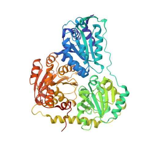
    4QPZ, 4QQ8

  • PubMed Abstract: 

    We describe a computationally designed enzyme, formolase (FLS), which catalyzes the carboligation of three one-carbon formaldehyde molecules into one three-carbon dihydroxyacetone molecule. The existence of FLS enables the design of a new carbon fixation pathway, the formolase pathway, consisting of a small number of thermodynamically favorable chemical transformations that convert formate into a three-carbon sugar in central metabolism. The formolase pathway is predicted to use carbon more efficiently and with less backward flux than any naturally occurring one-carbon assimilation pathway. When supplemented with enzymes carrying out the other steps in the pathway, FLS converts formate into dihydroxyacetone phosphate and other central metabolites in vitro. These results demonstrate how modern protein engineering and design tools can facilitate the construction of a completely new biosynthetic pathway.


  • Organizational Affiliation

    Department of Chemistry, Department of Biochemistry and Molecular Medicine, and Genome Center, University of California, Davis, CA 95616; Department of Biochemistry and the Institute for Protein Design, Biomolecular Structure and Design Program.


Macromolecules
Find similar proteins by:  (by identity cutoff)  |  3D Structure
Entity ID: 1
MoleculeChains Sequence LengthOrganismDetailsImage
Formolase
A, B, C, D
583Pseudomonas fluorescensMutation(s): 0 
Gene Names: bznB
UniProt
Find proteins for Q9F4L3 (Pseudomonas fluorescens)
Explore Q9F4L3 
Go to UniProtKB:  Q9F4L3
Entity Groups  
Sequence Clusters30% Identity50% Identity70% Identity90% Identity95% Identity100% Identity
UniProt GroupQ9F4L3
Sequence Annotations
Expand
  • Reference Sequence
Small Molecules
Ligands 3 Unique
IDChains Name / Formula / InChI Key2D Diagram3D Interactions
TPP
Query on TPP

Download Ideal Coordinates CCD File 
F [auth A],
J [auth B],
L [auth C],
O [auth D]
THIAMINE DIPHOSPHATE
C12 H19 N4 O7 P2 S
AYEKOFBPNLCAJY-UHFFFAOYSA-O
EDO
Query on EDO

Download Ideal Coordinates CCD File 
G [auth A],
H [auth A],
M [auth C],
P [auth D]
1,2-ETHANEDIOL
C2 H6 O2
LYCAIKOWRPUZTN-UHFFFAOYSA-N
MG
Query on MG

Download Ideal Coordinates CCD File 
E [auth A],
I [auth B],
K [auth C],
N [auth D]
MAGNESIUM ION
Mg
JLVVSXFLKOJNIY-UHFFFAOYSA-N
Experimental Data & Validation

Experimental Data

  • Method: X-RAY DIFFRACTION
  • Resolution: 2.88 Å
  • R-Value Free: 0.199 
  • R-Value Work: 0.169 
  • R-Value Observed: 0.171 
  • Space Group: P 43 21 2
Unit Cell:
Length ( Å )Angle ( ˚ )
a = 144.742α = 90
b = 144.742β = 90
c = 269.553γ = 90
Software Package:
Software NamePurpose
ADSCdata collection
PHASERphasing
REFMACrefinement
HKL-2000data reduction
HKL-2000data scaling

Structure Validation

View Full Validation Report


Ligand Structure Quality Assessment 


Entry History 

Deposition Data

Revision History  (Full details and data files)

  • Version 1.0: 2015-03-11
    Type: Initial release
  • Version 1.1: 2015-04-08
    Changes: Database references
  • Version 1.2: 2023-09-20
    Changes: Data collection, Database references, Derived calculations, Refinement description