4QLW

Azurin mutant M121E with iron


Experimental Data Snapshot

  • Method: X-RAY DIFFRACTION
  • Resolution: 2.00 Å
  • R-Value Free: 0.262 
  • R-Value Work: 0.205 
  • R-Value Observed: 0.207 

wwPDB Validation   3D Report Full Report


This is version 1.1 of the entry. See complete history


Literature

Redesigning the Blue Copper Azurin into a Redox-Active Mononuclear Nonheme Iron Protein: Preparation and Study of Fe(II)-M121E Azurin.

Liu, J.Meier, K.K.Tian, S.Zhang, J.L.Guo, H.Schulz, C.E.Robinson, H.Nilges, M.J.Munck, E.Lu, Y.

(2014) J Am Chem Soc 136: 12337-12344

  • DOI: https://doi.org/10.1021/ja505410u
  • Primary Citation of Related Structures:  
    4QKT, 4QLW

  • PubMed Abstract: 

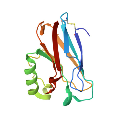
    Much progress has been made in designing heme and dinuclear nonheme iron enzymes. In contrast, engineering mononuclear nonheme iron enzymes is lagging, even though these enzymes belong to a large class that catalyzes quite diverse reactions. Herein we report spectroscopic and X-ray crystallographic studies of Fe(II)-M121E azurin (Az), by replacing the axial Met121 and Cu(II) in wild-type azurin (wtAz) with Glu and Fe(II), respectively. In contrast to the redox inactive Fe(II)-wtAz, the Fe(II)-M121EAz mutant can be readily oxidized by Na2IrCl6, and interestingly, the protein exhibits superoxide scavenging activity. Mössbauer and EPR spectroscopies, along with X-ray structural comparisons, revealed similarities and differences between Fe(II)-M121EAz, Fe(II)-wtAz, and superoxide reductase (SOR) and allowed design of the second generation mutant, Fe(II)-M121EM44KAz, that exhibits increased superoxide scavenging activity by 2 orders of magnitude. This finding demonstrates the importance of noncovalent secondary coordination sphere interactions in fine-tuning enzymatic activity.


  • Organizational Affiliation

    Department of Chemistry, University of Illinois at Urbana-Champaign , Urbana, Illinois 61801, United States.


Macromolecules
Find similar proteins by:  (by identity cutoff)  |  3D Structure
Entity ID: 1
MoleculeChains Sequence LengthOrganismDetailsImage
Azurin
A, B, C, D
128Pseudomonas aeruginosa PAO1Mutation(s): 1 
Gene Names: azuPA4922
UniProt
Find proteins for P00282 (Pseudomonas aeruginosa (strain ATCC 15692 / DSM 22644 / CIP 104116 / JCM 14847 / LMG 12228 / 1C / PRS 101 / PAO1))
Explore P00282 
Go to UniProtKB:  P00282
Entity Groups  
Sequence Clusters30% Identity50% Identity70% Identity90% Identity95% Identity100% Identity
UniProt GroupP00282
Sequence Annotations
Expand
  • Reference Sequence
Small Molecules
Ligands 3 Unique
IDChains Name / Formula / InChI Key2D Diagram3D Interactions
SO4
Query on SO4

Download Ideal Coordinates CCD File 
E [auth A],
F [auth A],
L [auth C]
SULFATE ION
O4 S
QAOWNCQODCNURD-UHFFFAOYSA-L
NO3
Query on NO3

Download Ideal Coordinates CCD File 
G [auth A]
H [auth A]
J [auth B]
K [auth B]
M [auth C]
G [auth A],
H [auth A],
J [auth B],
K [auth B],
M [auth C],
N [auth C],
O [auth C],
P [auth C],
R [auth D],
S [auth D]
NITRATE ION
N O3
NHNBFGGVMKEFGY-UHFFFAOYSA-N
FE
Query on FE

Download Ideal Coordinates CCD File 
I [auth B],
Q [auth D]
FE (III) ION
Fe
VTLYFUHAOXGGBS-UHFFFAOYSA-N
Experimental Data & Validation

Experimental Data

  • Method: X-RAY DIFFRACTION
  • Resolution: 2.00 Å
  • R-Value Free: 0.262 
  • R-Value Work: 0.205 
  • R-Value Observed: 0.207 
  • Space Group: P 21 21 21
Unit Cell:
Length ( Å )Angle ( ˚ )
a = 56.853α = 90
b = 80.208β = 90
c = 109.04γ = 90
Software Package:
Software NamePurpose
HKL-2000data collection
MOLREPphasing
REFMACrefinement
HKL-2000data reduction
HKL-2000data scaling

Structure Validation

View Full Validation Report


Entry History 

Deposition Data

Revision History  (Full details and data files)

  • Version 1.0: 2014-08-13
    Type: Initial release
  • Version 1.1: 2014-10-01
    Changes: Database references