4QLO

Crystal Structure of homoserine o-acetyltransferase from Staphylococcus aureus


Experimental Data Snapshot

  • Method: X-RAY DIFFRACTION
  • Resolution: 2.45 Å
  • R-Value Free: 0.291 
  • R-Value Work: 0.240 
  • R-Value Observed: 0.242 

wwPDB Validation   3D Report Full Report


This is version 1.2 of the entry. See complete history


Literature

Structure of homoserine O-acetyltransferase from Staphylococcus aureus: the first Gram-positive ortholog structure.

Thangavelu, B.Pavlovsky, A.G.Viola, R.

(2014) Acta Crystallogr Sect F Struct Biol Cryst Commun 70: 1340-1345

  • DOI: https://doi.org/10.1107/S2053230X14018664
  • Primary Citation of Related Structures:  
    4QLO

  • PubMed Abstract: 

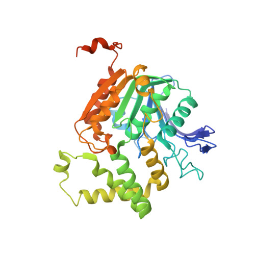
    Homoserine O-acetyltransferase (HTA) catalyzes the formation of L-O-acetyl-homoserine from L-homoserine through the transfer of an acetyl group from acetyl-CoA. This is the first committed step required for the biosynthesis of methionine in many fungi, Gram-positive bacteria and some Gram-negative bacteria. The structure of HTA from Staphylococcus aureus (SaHTA) has been determined to a resolution of 2.45 Å. The structure belongs to the α/β-hydrolase superfamily, consisting of two distinct domains: a core α/β-domain containing the catalytic site and a lid domain assembled into a helical bundle. The active site consists of a classical catalytic triad located at the end of a deep tunnel. Structure analysis revealed some important differences for SaHTA compared with the few known structures of HTA.


  • Organizational Affiliation

    Department of Chemistry and Biochemistry, The University of Toledo, Toledo, OH 43606, USA.


Macromolecules
Find similar proteins by:  (by identity cutoff)  |  3D Structure
Entity ID: 1
MoleculeChains Sequence LengthOrganismDetailsImage
homoserine O-acetyltransferase360Staphylococcus aureus subsp. aureus USA300_TCH1516Mutation(s): 0 
Gene Names: AZ30_00060metXSACOL0012USA300HOU_0012
EC: 2.3.1.31
Entity Groups  
Sequence Clusters30% Identity50% Identity70% Identity90% Identity95% Identity100% Identity
Sequence Annotations
Expand
  • Reference Sequence
Experimental Data & Validation

Experimental Data

  • Method: X-RAY DIFFRACTION
  • Resolution: 2.45 Å
  • R-Value Free: 0.291 
  • R-Value Work: 0.240 
  • R-Value Observed: 0.242 
  • Space Group: P 61 2 2
Unit Cell:
Length ( Å )Angle ( ˚ )
a = 49.155α = 90
b = 49.155β = 90
c = 481.417γ = 120
Software Package:
Software NamePurpose
CrystalCleardata collection
BALBESphasing
REFMACrefinement
HKL-2000data reduction
HKL-2000data scaling

Structure Validation

View Full Validation Report


Entry History 

Deposition Data

Revision History  (Full details and data files)

  • Version 1.0: 2014-08-20
    Type: Initial release
  • Version 1.1: 2014-12-17
    Changes: Database references
  • Version 1.2: 2024-02-28
    Changes: Data collection, Database references