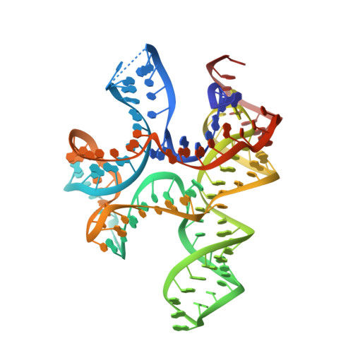
4QLN

structure of ydao riboswitch binding with c-di-dAMP


Experimental Data Snapshot

  • Method: X-RAY DIFFRACTION
  • Resolution: 2.65 Å
  • R-Value Free: 0.290 
  • R-Value Work: 0.217 
  • R-Value Observed: 0.221 

wwPDB Validation   3D Report Full Report


Ligand Structure Quality Assessment 


This is version 1.2 of the entry. See complete history


Literature

c-di-AMP binds the ydaO riboswitch in two pseudo-symmetry-related pockets.

Ren, A.Patel, D.J.

(2014) Nat Chem Biol 10: 780-786

  • DOI: https://doi.org/10.1038/nchembio.1606
  • Primary Citation of Related Structures:  
    4QLM, 4QLN

  • PubMed Abstract: 

    The ydaO riboswitch, involved in sporulation, osmotic stress responses and cell wall metabolism, targets the second messenger cyclic-di-AMP with subnanomolar affinity. We have solved the structure of c-di-AMP bound to the Thermoanaerobacter tengcongensis ydaO riboswitch, thereby identifying a five-helical scaffold containing a zippered-up bubble, a pseudoknot and long-range tertiary base pairs. Highlights include the identification of two c-di-AMP binding pockets on the same face of the riboswitch, related by pseudo-two-fold symmetry, with potential for cross-talk between sites mediated by adjacently positioned base-stacking alignments connecting pockets. The adenine rings of bound c-di-AMP molecules are wedged between bases and stabilized by stacking, base-sugar and sugar-sugar intermolecular hydrogen bonding interactions. The structural studies are complemented by isothermal titration calorimetry-based binding studies of mutants mediating key tertiary intermolecular contacts. The T. tengcongensis ydaO riboswitch, like its Bacillus subtilis counterpart, most likely functions through a transcription termination mechanism, with the c-di-AMP bound state representing an 'off' switch.


  • Organizational Affiliation

    Structural Biology Program, Memorial Sloan-Kettering Cancer Center, New York, New York, USA.


Macromolecules
Find similar nucleic acids by:  (by identity cutoff)  |  3D Structure
Entity ID: 1
MoleculeChains LengthOrganismImage
RNA (117-MER)125Caldanaerobacter subterraneus subsp. tengcongensis MB4
Sequence Annotations
Expand
  • Reference Sequence
Small Molecules
Ligands 2 Unique
IDChains Name / Formula / InChI Key2D Diagram3D Interactions
2BA
Query on 2BA

Download Ideal Coordinates CCD File 
B [auth A],
C [auth A]
(2R,3R,3aS,5R,7aR,9R,10R,10aS,12R,14aR)-2,9-bis(6-amino-9H-purin-9-yl)octahydro-2H,7H-difuro[3,2-d:3',2'-j][1,3,7,9,2,8 ]tetraoxadiphosphacyclododecine-3,5,10,12-tetrol 5,12-dioxide
C20 H24 N10 O12 P2
PDXMFTWFFKBFIN-XPWFQUROSA-N
MG
Query on MG

Download Ideal Coordinates CCD File 
D [auth A],
E [auth A]
MAGNESIUM ION
Mg
JLVVSXFLKOJNIY-UHFFFAOYSA-N
Experimental Data & Validation

Experimental Data

  • Method: X-RAY DIFFRACTION
  • Resolution: 2.65 Å
  • R-Value Free: 0.290 
  • R-Value Work: 0.217 
  • R-Value Observed: 0.221 
  • Space Group: C 2 2 21
Unit Cell:
Length ( Å )Angle ( ˚ )
a = 77.874α = 90
b = 87.051β = 90
c = 144.553γ = 90
Software Package:
Software NamePurpose
XDSdata scaling
PHASERphasing
PHENIXrefinement
XDSdata reduction

Structure Validation

View Full Validation Report


Ligand Structure Quality Assessment 


Entry History 

Deposition Data

Revision History  (Full details and data files)

  • Version 1.0: 2014-08-13
    Type: Initial release
  • Version 1.1: 2014-09-03
    Changes: Database references
  • Version 1.2: 2024-02-28
    Changes: Data collection, Database references, Derived calculations, Structure summary