4QK8

Thermoanaerobacter pseudethanolicus c-di-AMP riboswitch

  • Classification: RNA
  • Mutation(s): No 

  • Deposited: 2014-06-05 Released: 2014-08-06 
  • Deposition Author(s): Gao, A., Serganov, A.

Experimental Data Snapshot

  • Method: X-RAY DIFFRACTION
  • Resolution: 3.05 Å
  • R-Value Free: 0.195 
  • R-Value Work: 0.181 
  • R-Value Observed: 0.183 

wwPDB Validation   3D Report Full Report


Ligand Structure Quality Assessment 


This is version 1.3 of the entry. See complete history


Literature

Structural insights into recognition of c-di-AMP by the ydaO riboswitch.

Gao, A.Serganov, A.

(2014) Nat Chem Biol 10: 787-792

  • DOI: https://doi.org/10.1038/nchembio.1607
  • Primary Citation of Related Structures:  
    4QK8, 4QK9, 4QKA

  • PubMed Abstract: 

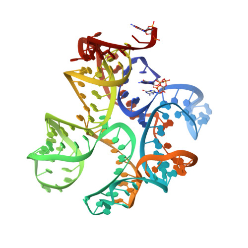
    Bacterial second messenger cyclic di-AMP (c-di-AMP) is implicated in signaling DNA damage and cell wall stress through interactions with several protein receptors and a widespread ydaO-type riboswitch. We report the crystal structures of c-di-AMP riboswitches from Thermoanaerobacter pseudethanolicus and Thermovirga lienii determined at ∼3.0-Å resolution. In both species, the RNA adopts an unforeseen 'square'-shaped pseudosymmetrical architecture that features two three-way junctions, a turn and a pseudoknot, positioned in the square corners. Uncharacteristically for riboswitches, the structure is stapled by two ligand molecules that span the interior of the structure and employ similar noncanonical interactions for RNA recognition. Mutations in either ligand-binding site negatively affect c-di-AMP binding, suggesting that the riboswitch-triggered genetic response requires contribution of both ligands. Our data provide what are to our knowledge the first insights into specific sensing of c-di-AMP and a molecular mechanism underlying the common c-di-AMP-dependent control of essential cellular processes in bacteria.


  • Organizational Affiliation

    Department of Biochemistry and Molecular Pharmacology, New York University School of Medicine, New York, New York, USA.


Macromolecules
Find similar nucleic acids by:  (by identity cutoff)  |  3D Structure
Entity ID: 1
MoleculeChains LengthOrganismImage
C-di-AMP riboswitch122N/A
Sequence Annotations
Expand
  • Reference Sequence
Small Molecules
Ligands 4 Unique
IDChains Name / Formula / InChI Key2D Diagram3D Interactions
2BA
Query on 2BA

Download Ideal Coordinates CCD File 
B [auth A],
C [auth A]
(2R,3R,3aS,5R,7aR,9R,10R,10aS,12R,14aR)-2,9-bis(6-amino-9H-purin-9-yl)octahydro-2H,7H-difuro[3,2-d:3',2'-j][1,3,7,9,2,8 ]tetraoxadiphosphacyclododecine-3,5,10,12-tetrol 5,12-dioxide
C20 H24 N10 O12 P2
PDXMFTWFFKBFIN-XPWFQUROSA-N
SO4
Query on SO4

Download Ideal Coordinates CCD File 
I [auth A]SULFATE ION
O4 S
QAOWNCQODCNURD-UHFFFAOYSA-L
K
Query on K

Download Ideal Coordinates CCD File 
F [auth A]POTASSIUM ION
K
NPYPAHLBTDXSSS-UHFFFAOYSA-N
MG
Query on MG

Download Ideal Coordinates CCD File 
D [auth A],
E [auth A],
G [auth A],
H [auth A]
MAGNESIUM ION
Mg
JLVVSXFLKOJNIY-UHFFFAOYSA-N
Experimental Data & Validation

Experimental Data

  • Method: X-RAY DIFFRACTION
  • Resolution: 3.05 Å
  • R-Value Free: 0.195 
  • R-Value Work: 0.181 
  • R-Value Observed: 0.183 
  • Space Group: P 31 2 1
Unit Cell:
Length ( Å )Angle ( ˚ )
a = 114.868α = 90
b = 114.868β = 90
c = 114.709γ = 120
Software Package:
Software NamePurpose
ADSCdata collection
PHENIXmodel building
PHENIXrefinement
HKL-2000data reduction
HKL-2000data scaling
PHENIXphasing

Structure Validation

View Full Validation Report


Ligand Structure Quality Assessment 


Entry History 

Deposition Data

Revision History  (Full details and data files)

  • Version 1.0: 2014-08-06
    Type: Initial release
  • Version 1.1: 2014-08-27
    Changes: Database references
  • Version 1.2: 2014-09-10
    Changes: Database references
  • Version 1.3: 2024-02-28
    Changes: Data collection, Database references, Derived calculations, Structure summary