4QF9

Structure of GluK1 ligand-binding domain (S1S2) in complex with (S)-2-amino-4-(2,3-dioxo-1,2,3,4-tetrahydroquinoxalin-6-yl)butanoic acid at 2.28 A resolution


Experimental Data Snapshot

  • Method: X-RAY DIFFRACTION
  • Resolution: 2.28 Å
  • R-Value Free: 0.246 
  • R-Value Work: 0.198 
  • R-Value Observed: 0.200 

wwPDB Validation   3D Report Full Report


Ligand Structure Quality Assessment 


This is version 1.4 of the entry. See complete history


Literature

Binding Mode of an alpha-Amino Acid-Linked Quinoxaline-2,3-dione Analogue at Glutamate Receptor Subtype GluK1.

Demmer, C.S.Moller, C.Brown, P.M.Han, L.Pickering, D.S.Nielsen, B.Bowie, D.Frydenvang, K.Kastrup, J.S.Bunch, L.

(2015) ACS Chem Neurosci 6: 845-854

  • DOI: https://doi.org/10.1021/acschemneuro.5b00038
  • Primary Citation of Related Structures:  
    4QF9

  • PubMed Abstract: 

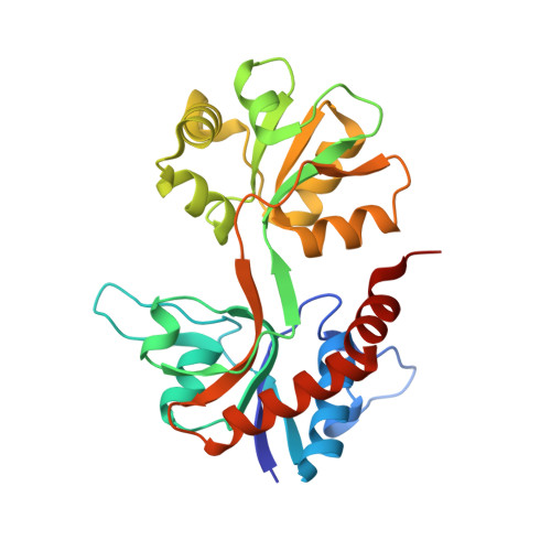
    Two α-amino acid-functionalized quinoxalines, 1a (CNG-10301) and 1b (CNG-10300), of a quinoxaline moiety coupled to an amino acid moiety were designed, synthesized, and characterized pharmacologically. While 1a displayed low affinity at native AMPA, KA, and NMDA receptors, and at homomeric GluK1,3 receptors, the affinity for GluK2 was in the midmicromolar range (Ki = 136 μM), 1b displayed low to midmicromolar range binding affinity at all the iGluRs (Ki = 9-126 μM). In functional experiments (outside-out patches excised from transfected HEK293T cells), 100 μM 1a partially blocked GluK1 (33% peak response), while GluK2 was unaffected (96% peak response). Furthermore, 1a was shown not to be an agonist at GluK1 and GluK2 at 100 μM. On the other hand, 100 μM 1b fully antagonized GluK1 (8% peak response) but only partially blocked GluK2 (33% peak response). An X-ray structure at 2.3 Å resolution of 1b in the GluK1-LBD (ligand-binding domain) disclosed an unexpected binding mode compared to the predictions made during the design phase; the quinoxaline moiety remains to act as an amino acid bioisostere, but the amino acid moiety is oriented into a new area within the GluK1 receptor. The structure of the GluK1-LBD with 1b showed a large variation in domain openings of the three molecules from 25° to 49°, demonstrating that the GluK1-LBD is capable of undergoing major domain movements.


Macromolecules
Find similar proteins by:  (by identity cutoff)  |  3D Structure
Entity ID: 1
MoleculeChains Sequence LengthOrganismDetailsImage
Glutamate receptor ionotropic, kainate 1
A, B, C
257Rattus norvegicusMutation(s): 0 
Gene Names: Glur5Grik1
UniProt
Find proteins for P22756 (Rattus norvegicus)
Explore P22756 
Go to UniProtKB:  P22756
Entity Groups  
Sequence Clusters30% Identity50% Identity70% Identity90% Identity95% Identity100% Identity
UniProt GroupP22756
Sequence Annotations
Expand
  • Reference Sequence
Small Molecules
Ligands 6 Unique
IDChains Name / Formula / InChI Key2D Diagram3D Interactions
35K
Query on 35K

Download Ideal Coordinates CCD File 
D [auth A],
H [auth B],
O [auth C]
(2S)-2-amino-4-(2,3-dioxo-1,2,3,4-tetrahydroquinoxalin-6-yl)butanoic acid
C12 H13 N3 O4
HOBORVHHDCBXLX-ZETCQYMHSA-N
PG4
Query on PG4

Download Ideal Coordinates CCD File 
F [auth A],
N [auth B]
TETRAETHYLENE GLYCOL
C8 H18 O5
UWHCKJMYHZGTIT-UHFFFAOYSA-N
SO4
Query on SO4

Download Ideal Coordinates CCD File 
E [auth A],
I [auth B],
J [auth B],
P [auth C]
SULFATE ION
O4 S
QAOWNCQODCNURD-UHFFFAOYSA-L
GOL
Query on GOL

Download Ideal Coordinates CCD File 
K [auth B],
L [auth B],
M [auth B]
GLYCEROL
C3 H8 O3
PEDCQBHIVMGVHV-UHFFFAOYSA-N
ACT
Query on ACT

Download Ideal Coordinates CCD File 
Q [auth C]ACETATE ION
C2 H3 O2
QTBSBXVTEAMEQO-UHFFFAOYSA-M
CL
Query on CL

Download Ideal Coordinates CCD File 
G [auth A]CHLORIDE ION
Cl
VEXZGXHMUGYJMC-UHFFFAOYSA-M
Binding Affinity Annotations 
IDSourceBinding Affinity
35K Binding MOAD:  4QF9 Ki: 3.71e+4 (nM) from 1 assay(s)
Experimental Data & Validation

Experimental Data

  • Method: X-RAY DIFFRACTION
  • Resolution: 2.28 Å
  • R-Value Free: 0.246 
  • R-Value Work: 0.198 
  • R-Value Observed: 0.200 
  • Space Group: C 1 2 1
Unit Cell:
Length ( Å )Angle ( ˚ )
a = 117.265α = 90
b = 86.802β = 111.62
c = 78.282γ = 90
Software Package:
Software NamePurpose
SCALAdata scaling
PHASERphasing
PHENIXrefinement
PDB_EXTRACTdata extraction
XDSdata reduction

Structure Validation

View Full Validation Report


Ligand Structure Quality Assessment 


Entry History 

Deposition Data

Revision History  (Full details and data files)

  • Version 1.0: 2015-04-22
    Type: Initial release
  • Version 1.1: 2015-07-01
    Changes: Data collection, Database references
  • Version 1.2: 2017-07-26
    Changes: Data collection, Source and taxonomy
  • Version 1.3: 2017-08-09
    Changes: Database references
  • Version 1.4: 2023-09-20
    Changes: Data collection, Database references, Derived calculations, Refinement description