4QDJ

Crystal structure of magnesium protoporphyrin IX methyltransferase (ChlM) from Synechocystis PCC 6803 with bound SAM


Experimental Data Snapshot

  • Method: X-RAY DIFFRACTION
  • Resolution: 1.60 Å
  • R-Value Free: 0.180 
  • R-Value Work: 0.141 
  • R-Value Observed: 0.143 

wwPDB Validation   3D Report Full Report


Ligand Structure Quality Assessment 


This is version 1.2 of the entry. See complete history


Literature

Structural insights into the catalytic mechanism of Synechocystis magnesium protoporphyrin IX O-methyltransferase (ChlM).

Chen, X.Wang, X.Feng, J.Chen, Y.Fang, Y.Zhao, S.Zhao, A.Zhang, M.Liu, L.

(2014) J Biol Chem 289: 25690-25698

  • DOI: https://doi.org/10.1074/jbc.M114.584920
  • Primary Citation of Related Structures:  
    4QDJ, 4QDK

  • PubMed Abstract: 

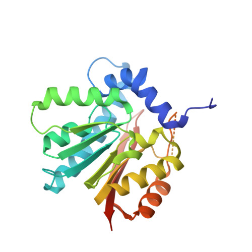
    Magnesium protoporphyrin IX O-methyltransferase (ChlM) catalyzes transfer of the methyl group from S-adenosylmethionine to the carboxyl group of the C13 propionate side chain of magnesium protoporphyrin IX. This reaction is the second committed step in chlorophyll biosynthesis from protoporphyrin IX. Here we report the crystal structures of ChlM from the cyanobacterium Synechocystis sp. PCC 6803 in complex with S-adenosylmethionine and S-adenosylhomocysteine at resolutions of 1.6 and 1.7 Å, respectively. The structures illustrate the molecular basis for cofactor and substrate binding and suggest that conformational changes of the two "arm" regions may modulate binding and release of substrates/products to and from the active site. Tyr-28 and His-139 were identified to play essential roles for methyl transfer reaction but are not indispensable for cofactor/substrate binding. Based on these structural and functional findings, a catalytic model is proposed.


  • Organizational Affiliation

    From the Photosynthesis Research Center, Key Laboratory of Photobiology, Institute of Botany, Chinese Academy of Sciences, Beijing, 100093, China, the University of Chinese Academy of Sciences, Beijing, 100049, China.


Macromolecules
Find similar proteins by:  (by identity cutoff)  |  3D Structure
Entity ID: 1
MoleculeChains Sequence LengthOrganismDetailsImage
Magnesium-protoporphyrin O-methyltransferase238Synechocystis sp. PCC 6803 substr. KazusaMutation(s): 0 
Gene Names: chlMslr0525
EC: 2.1.1.11
UniProt
Find proteins for Q55467 (Synechocystis sp. (strain PCC 6803 / Kazusa))
Explore Q55467 
Go to UniProtKB:  Q55467
Entity Groups  
Sequence Clusters30% Identity50% Identity70% Identity90% Identity95% Identity100% Identity
UniProt GroupQ55467
Sequence Annotations
Expand
  • Reference Sequence
Small Molecules
Ligands 2 Unique
IDChains Name / Formula / InChI Key2D Diagram3D Interactions
SAM
Query on SAM

Download Ideal Coordinates CCD File 
B [auth A]S-ADENOSYLMETHIONINE
C15 H22 N6 O5 S
MEFKEPWMEQBLKI-FCKMPRQPSA-N
GOL
Query on GOL

Download Ideal Coordinates CCD File 
C [auth A]GLYCEROL
C3 H8 O3
PEDCQBHIVMGVHV-UHFFFAOYSA-N
Experimental Data & Validation

Experimental Data

  • Method: X-RAY DIFFRACTION
  • Resolution: 1.60 Å
  • R-Value Free: 0.180 
  • R-Value Work: 0.141 
  • R-Value Observed: 0.143 
  • Space Group: P 21 21 21
Unit Cell:
Length ( Å )Angle ( ˚ )
a = 49.319α = 90
b = 61.026β = 90
c = 68.892γ = 90
Software Package:
Software NamePurpose
ADSCdata collection
PHENIXmodel building
PHENIXrefinement
HKL-2000data reduction
HKL-2000data scaling
PHENIXphasing

Structure Validation

View Full Validation Report


Ligand Structure Quality Assessment 


Entry History 

Deposition Data

Revision History  (Full details and data files)

  • Version 1.0: 2014-08-06
    Type: Initial release
  • Version 1.1: 2015-08-19
    Changes: Database references
  • Version 1.2: 2024-03-20
    Changes: Data collection, Database references, Derived calculations