


4QAB
X-RAY STRUCTURE of ACETYLCHOLINE BINDING PROTEIN (ACHBP) IN COMPLEX WITH 4-(MORPHOLIN-4-YL)-6-[4-(TRIFLUOROMETHYL)PHENYL]PYRIMIDIN-2-AMINE
- PDB DOI: https://doi.org/10.2210/pdb4QAB/pdb
- Classification: Acetylcholine-Binding Protein
- Organism(s): Lymnaea stagnalis
- Expression System: Homo sapiens
- Mutation(s): No
- Deposited: 2014-05-03 Released: 2014-07-16
Experimental Data Snapshot
- Method: X-RAY DIFFRACTION
- Resolution: 2.98 Å
- R-Value Free: 0.233
- R-Value Work: 0.150
- R-Value Observed: 0.153

This is version 1.4 of the entry. See complete history.

Macromolecules
Find similar proteins by:
(by identity cutoff) | 3D Structure
Entity ID: 1 | |||||
|---|---|---|---|---|---|
| Molecule | Chains | Sequence Length | Organism | Details | Image |
| Acetylcholine-binding protein | 217 | Lymnaea stagnalis | Mutation(s): 0 | 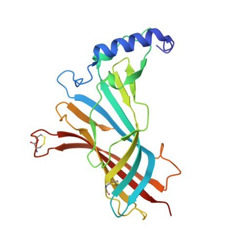 | |
UniProt | |||||
Find proteins for P58154 (Lymnaea stagnalis) Explore P58154 Go to UniProtKB: P58154 | |||||
Entity Groups | |||||
| Sequence Clusters | 30% Identity50% Identity70% Identity90% Identity95% Identity100% Identity | ||||
| UniProt Group | P58154 | ||||
Sequence AnnotationsExpand | |||||
| |||||
Small Molecules
| Ligands 3 Unique | |||||
|---|---|---|---|---|---|
| ID | Chains | Name / Formula / InChI Key | 2D Diagram | 3D Interactions | |
| KK2 Query on KK2 | BA [auth G] EA [auth H] HA [auth I] K [auth A] KA [auth J] | 4-(morpholin-4-yl)-6-[4-(trifluoromethyl)phenyl]pyrimidin-2-amine C15 H15 F3 N4 O XZLXJMNYYMKDDT-UHFFFAOYSA-N |  | ||
| NAG Query on NAG | CA [auth G] FA [auth H] IA [auth I] L [auth A] LA [auth J] | 2-acetamido-2-deoxy-beta-D-glucopyranose C8 H15 N O6 OVRNDRQMDRJTHS-FMDGEEDCSA-N |  | ||
| PO4 Query on PO4 | AA [auth F] DA [auth G] GA [auth H] JA [auth I] M [auth A] | PHOSPHATE ION O4 P NBIIXXVUZAFLBC-UHFFFAOYSA-K |  | ||
Experimental Data & Validation
Experimental Data
- Method: X-RAY DIFFRACTION
- Resolution: 2.98 Å
- R-Value Free: 0.233
- R-Value Work: 0.150
- R-Value Observed: 0.153
- Space Group: P 21 21 21
Unit Cell:
| Length ( Å ) | Angle ( ˚ ) |
|---|---|
| a = 122.639 | α = 90 |
| b = 134.015 | β = 90 |
| c = 147.02 | γ = 90 |
| Software Name | Purpose |
|---|---|
| HKL-2000 | data collection |
| PHASER | phasing |
| PHENIX | refinement |
| HKL-2000 | data reduction |
| HKL-2000 | data scaling |
Entry History
Deposition Data
- Released Date: 2014-07-16 Deposition Author(s): Kaczanowska, K., Harel, M., Radic, Z., Changeux, J.-P., Finn, M.G., Taylor, P.
Revision History (Full details and data files)
- Version 1.0: 2014-07-16
Type: Initial release - Version 1.1: 2014-07-30
Changes: Database references - Version 1.2: 2014-08-13
Changes: Database references - Version 1.3: 2018-01-24
Changes: Structure summary - Version 1.4: 2020-07-29
Type: Remediation
Reason: Carbohydrate remediation
Changes: Data collection, Database references, Derived calculations, Structure summary
