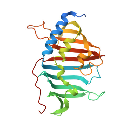
4QA8

Crystal structure of LprF from Mycobacterium bovis


Experimental Data Snapshot

  • Method: X-RAY DIFFRACTION
  • Resolution: 1.10 Å
  • R-Value Free: 0.189 
  • R-Value Work: 0.164 
  • R-Value Observed: 0.165 

wwPDB Validation   3D Report Full Report


Ligand Structure Quality Assessment 


This is version 1.1 of the entry. See complete history


Literature

Crystal structure and functional implications of LprF from Mycobacterium tuberculosis and M. bovis

Kim, J.S.Jiao, L.Oh, J.I.Ha, N.C.Kim, Y.H.

(2014) Acta Crystallogr D Biol Crystallogr 70: 2619-2630

  • DOI: https://doi.org/10.1107/S1399004714016599
  • Primary Citation of Related Structures:  
    4QA8

  • PubMed Abstract: 

    The Gram-positive bacteria Mycobacterium tuberculosis and M. bovis are causative agents of tuberculosis in humans and cattle. The lipoprotein LprF is found in M. tuberculosis and M. bovis but not in the nonpathogenic M. smegmatis. To date, the role of LprF remains to be elucidated. In this study, the crystal structure of LprF has been determined at 1.1 Å resolution. The overall structure is similar to that of a homologue, LprG, with a central hydrophobic cavity that binds a triacylated glycolipid. LprF exhibited a central cavity structure similar to that of LprG, but with a smaller cavity that binds two alkyl chains. Consistently, subsequent mass-spectrometric analysis revealed that the bound ligand was a diacylated glycolipid, as found in the structure. Furthermore, an increased ratio of lipoarabinomannan to lipomannan in the mycobacterial cell wall was observed when lprF was introduced into M. smegmatis. These observations suggested that LprF transfers the diacylated glycolipid from the plasma membrane to the cell wall, which might be related to the pathogenesis of the bacteria.


  • Organizational Affiliation

    College of Pharmacy and Research Institute for Drug Development, Pusan National University, Busan, Republic of Korea.


Macromolecules
Find similar proteins by:  (by identity cutoff)  |  3D Structure
Entity ID: 1
MoleculeChains Sequence LengthOrganismDetailsImage
Putative lipoprotein LprF229Mycobacterium tuberculosis variant bovis AF2122/97Mutation(s): 0 
Gene Names: lprFMb1403
UniProt
Find proteins for P65315 (Mycobacterium bovis (strain ATCC BAA-935 / AF2122/97))
Explore P65315 
Go to UniProtKB:  P65315
Entity Groups  
Sequence Clusters30% Identity50% Identity70% Identity90% Identity95% Identity100% Identity
UniProt GroupP65315
Sequence Annotations
Expand
  • Reference Sequence
Small Molecules
Ligands 1 Unique
IDChains Name / Formula / InChI Key2D Diagram3D Interactions
PJZ
Query on PJZ

Download Ideal Coordinates CCD File 
B [auth A](2R)-2-(dodecanoyloxy)propyl (4E,6E,8E,10E,12E)-pentadeca-4,6,8,10,12-pentaenoate
C30 H48 O4
ONKSLGBBXPETKB-WGIZECGSSA-N
Experimental Data & Validation

Experimental Data

  • Method: X-RAY DIFFRACTION
  • Resolution: 1.10 Å
  • R-Value Free: 0.189 
  • R-Value Work: 0.164 
  • R-Value Observed: 0.165 
  • Space Group: P 1
Unit Cell:
Length ( Å )Angle ( ˚ )
a = 31.514α = 76.4
b = 32.381β = 80.79
c = 51.717γ = 71.23
Software Package:
Software NamePurpose
HKL-2000data collection
MOLREPphasing
PHENIXrefinement
DENZOdata reduction
HKL-2000data scaling

Structure Validation

View Full Validation Report


Ligand Structure Quality Assessment 


Entry History 

Deposition Data

Revision History  (Full details and data files)

  • Version 1.0: 2014-10-22
    Type: Initial release
  • Version 1.1: 2024-03-20
    Changes: Data collection, Database references, Derived calculations