4Q67

Staphylococcus aureus F98Y mutant dihydrofolate reductase complexed with NADPH


Experimental Data Snapshot

  • Method: X-RAY DIFFRACTION
  • Resolution: 2.04 Å
  • R-Value Free: 0.233 
  • R-Value Work: 0.175 
  • R-Value Observed: 0.178 

wwPDB Validation   3D Report Full Report


Ligand Structure Quality Assessment 


This is version 1.2 of the entry. See complete history


Literature

Protein design algorithms predict viable resistance to an experimental antifolate.

Reeve, S.M.Gainza, P.Frey, K.M.Georgiev, I.Donald, B.R.Anderson, A.C.

(2015) Proc Natl Acad Sci U S A 112: 749-754

  • DOI: https://doi.org/10.1073/pnas.1411548112
  • Primary Citation of Related Structures:  
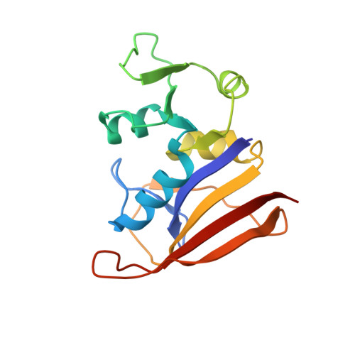
    4Q67, 4Q6A

  • PubMed Abstract: 

    Methods to accurately predict potential drug target mutations in response to early-stage leads could drive the design of more resilient first generation drug candidates. In this study, a structure-based protein design algorithm (K* in the OSPREY suite) was used to prospectively identify single-nucleotide polymorphisms that confer resistance to an experimental inhibitor effective against dihydrofolate reductase (DHFR) from Staphylococcus aureus. Four of the top-ranked mutations in DHFR were found to be catalytically competent and resistant to the inhibitor. Selection of resistant bacteria in vitro reveals that two of the predicted mutations arise in the background of a compensatory mutation. Using enzyme kinetics, microbiology, and crystal structures of the complexes, we determined the fitness of the mutant enzymes and strains, the structural basis of resistance, and the compensatory relationship of the mutations. To our knowledge, this work illustrates the first application of protein design algorithms to prospectively predict viable resistance mutations that arise in bacteria under antibiotic pressure.


  • Organizational Affiliation

    Department of Pharmaceutical Sciences, University of Connecticut, Storrs, CT 06269; and.


Macromolecules
Find similar proteins by:  (by identity cutoff)  |  3D Structure
Entity ID: 1
MoleculeChains Sequence LengthOrganismDetailsImage
Dihydrofolate reductase160Staphylococcus aureus MUF168Mutation(s): 0 
Gene Names: dfrBSaDHFRY000_11620
EC: 1.5.1.3
Entity Groups  
Sequence Clusters30% Identity50% Identity70% Identity90% Identity95% Identity100% Identity
Sequence Annotations
Expand
  • Reference Sequence
Small Molecules
Ligands 1 Unique
IDChains Name / Formula / InChI Key2D Diagram3D Interactions
NAP
Query on NAP

Download Ideal Coordinates CCD File 
B [auth A]NADP NICOTINAMIDE-ADENINE-DINUCLEOTIDE PHOSPHATE
C21 H28 N7 O17 P3
XJLXINKUBYWONI-NNYOXOHSSA-N
Experimental Data & Validation

Experimental Data

  • Method: X-RAY DIFFRACTION
  • Resolution: 2.04 Å
  • R-Value Free: 0.233 
  • R-Value Work: 0.175 
  • R-Value Observed: 0.178 
  • Space Group: P 61 2 2
Unit Cell:
Length ( Å )Angle ( ˚ )
a = 79.26α = 90
b = 79.26β = 90
c = 107.42γ = 120
Software Package:
Software NamePurpose
StructureStudiodata collection
PHASERphasing
PHENIXrefinement

Structure Validation

View Full Validation Report


Ligand Structure Quality Assessment 


Entry History 

Deposition Data

Revision History  (Full details and data files)

  • Version 1.0: 2014-12-31
    Type: Initial release
  • Version 1.1: 2015-02-18
    Changes: Database references
  • Version 1.2: 2023-09-20
    Changes: Data collection, Database references, Derived calculations, Refinement description