4Q4E

Crystal structure of E.coli aminopeptidase N in complex with actinonin


Experimental Data Snapshot

  • Method: X-RAY DIFFRACTION
  • Resolution: 1.90 Å
  • R-Value Free: 0.181 
  • R-Value Work: 0.147 
  • R-Value Observed: 0.149 

wwPDB Validation   3D Report Full Report


Ligand Structure Quality Assessment 


This is version 1.2 of the entry. See complete history


Literature

Structural basis for the inhibition of M1 family aminopeptidases by the natural product actinonin: Crystal structure in complex with E. coli aminopeptidase N.

Ganji, R.J.Reddi, R.Gumpena, R.Marapaka, A.K.Arya, T.Sankoju, P.Bhukya, S.Addlagatta, A.

(2015) Protein Sci 24: 823-831

  • DOI: https://doi.org/10.1002/pro.2653
  • Primary Citation of Related Structures:  
    4Q4E, 4Q4I

  • PubMed Abstract: 

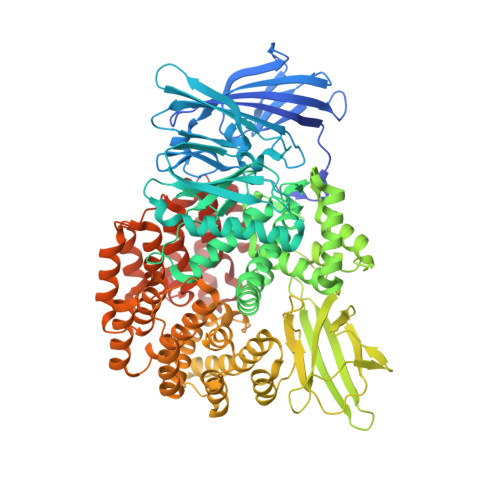
    Actinonin is a pseudotripeptide that displays a high affinity towards metalloproteases including peptide deformylases (PDFs) and M1 family aminopeptidases. PDF and M1 family aminopeptidases belong to thermolysin-metzincin superfamily. One of the major differences in terms of substrate binding pockets between these families is presence (in M1 aminopeptidases) or absence (in PDFs) of an S1 substrate pocket. The binding mode of actinonin to PDFs has been established previously; however, it is not clear how the actinonin, without a P1 residue, would bind to the M1 aminopeptidases. Here we describe the crystal structure of Escherichia coli aminopeptidase N (ePepN), a model protein of the M1 family aminopeptidases in complex with actinonin. For comparison we have also determined the structure of ePepN in complex with a well-known tetrapeptide inhibitor, amastatin. From the comparison of the actinonin and amastatin ePepN complexes, it is clear that the P1 residue is not critical as long as strong metal chelating head groups, like hydroxamic acid or α-hydroxy ketone, are present. Results from this study will be useful for the design of selective and efficient hydroxamate inhibitors against M1 family aminopeptidases.


  • Organizational Affiliation

    Center for Chemical Biology, CSIR-Indian Institute of Chemical Technology, Hyderabad, 500007, Telangana, India.


Macromolecules
Find similar proteins by:  (by identity cutoff)  |  3D Structure
Entity ID: 1
MoleculeChains Sequence LengthOrganismDetailsImage
Aminopeptidase N891Escherichia coli K-12Mutation(s): 0 
Gene Names: pepNb0932JW0915
EC: 3.4.11.2
UniProt
Find proteins for P04825 (Escherichia coli (strain K12))
Explore P04825 
Go to UniProtKB:  P04825
Entity Groups  
Sequence Clusters30% Identity50% Identity70% Identity90% Identity95% Identity100% Identity
UniProt GroupP04825
Sequence Annotations
Expand
  • Reference Sequence
Small Molecules
Ligands 4 Unique
IDChains Name / Formula / InChI Key2D Diagram3D Interactions
BB2
Query on BB2

Download Ideal Coordinates CCD File 
C [auth A]ACTINONIN
C19 H35 N3 O5
XJLATMLVMSFZBN-VYDXJSESSA-N
GOL
Query on GOL

Download Ideal Coordinates CCD File 
D [auth A]
E [auth A]
F [auth A]
G [auth A]
H [auth A]
D [auth A],
E [auth A],
F [auth A],
G [auth A],
H [auth A],
I [auth A],
J [auth A],
K [auth A]
GLYCEROL
C3 H8 O3
PEDCQBHIVMGVHV-UHFFFAOYSA-N
ZN
Query on ZN

Download Ideal Coordinates CCD File 
B [auth A]ZINC ION
Zn
PTFCDOFLOPIGGS-UHFFFAOYSA-N
NA
Query on NA

Download Ideal Coordinates CCD File 
L [auth A]SODIUM ION
Na
FKNQFGJONOIPTF-UHFFFAOYSA-N
Binding Affinity Annotations 
IDSourceBinding Affinity
BB2 Binding MOAD:  4Q4E IC50: 190 (nM) from 1 assay(s)
Experimental Data & Validation

Experimental Data

  • Method: X-RAY DIFFRACTION
  • Resolution: 1.90 Å
  • R-Value Free: 0.181 
  • R-Value Work: 0.147 
  • R-Value Observed: 0.149 
  • Space Group: P 31 2 1
Unit Cell:
Length ( Å )Angle ( ˚ )
a = 120.45α = 90
b = 120.45β = 90
c = 170.748γ = 120
Software Package:
Software NamePurpose
HKL-3000data collection
MOLREPphasing
REFMACrefinement
HKL-3000data reduction
HKL-3000data scaling

Structure Validation

View Full Validation Report


Ligand Structure Quality Assessment 


Entry History 

Deposition Data

Revision History  (Full details and data files)

  • Version 1.0: 2015-04-15
    Type: Initial release
  • Version 1.1: 2019-12-04
    Changes: Data collection, Database references
  • Version 1.2: 2022-08-24
    Changes: Database references, Derived calculations