


4PXF
Crystal structure of the active G-protein-coupled receptor opsin in complex with the finger-loop peptide derived from the full-length arrestin-1
- PDB DOI: https://doi.org/10.2210/pdb4PXF/pdb
- Classification: SIGNALING PROTEIN
- Organism(s): Bos taurus
- Mutation(s): No
- Membrane Protein: Yes OPMPDBTMMemProtMDmpstruc
- Deposited: 2014-03-23 Released: 2014-09-17
Experimental Data Snapshot
- Method: X-RAY DIFFRACTION
- Resolution: 2.75 Å
- R-Value Free: 0.251
- R-Value Work: 0.215
- R-Value Observed: 0.217

This is version 2.1 of the entry. See complete history.

Macromolecules
Find similar proteins by:
(by identity cutoff) | 3D Structure
Entity ID: 1 | |||||
|---|---|---|---|---|---|
| Molecule | Chains | Sequence Length | Organism | Details | Image |
| Rhodopsin | 348 | Bos taurus | Mutation(s): 0 Membrane Entity: Yes |  | |
UniProt | |||||
Find proteins for P02699 (Bos taurus) Explore P02699 Go to UniProtKB: P02699 | |||||
Entity Groups | |||||
| Sequence Clusters | 30% Identity50% Identity70% Identity90% Identity95% Identity100% Identity | ||||
| UniProt Group | P02699 | ||||
Sequence AnnotationsExpand | |||||
| |||||
Find similar proteins by: Sequence | 3D Structure
Entity ID: 2 | |||||
|---|---|---|---|---|---|
| Molecule | Chains | Sequence Length | Organism | Details | Image |
| S-arrestin | 11 | Bos taurus | Mutation(s): 0 Membrane Entity: Yes | 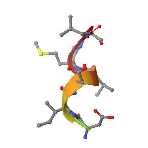 | |
UniProt | |||||
Find proteins for P08168 (Bos taurus) Explore P08168 Go to UniProtKB: P08168 | |||||
Entity Groups | |||||
| Sequence Clusters | 30% Identity50% Identity70% Identity90% Identity95% Identity100% Identity | ||||
| UniProt Group | P08168 | ||||
Sequence AnnotationsExpand | |||||
| |||||
Oligosaccharides
Entity ID: 3 | |||||
|---|---|---|---|---|---|
| Molecule | Chains | Length | 2D Diagram | Glycosylation | 3D Interactions |
| alpha-D-mannopyranose-(1-3)-beta-D-mannopyranose-(1-4)-2-acetamido-2-deoxy-beta-D-glucopyranose-(1-4)-2-acetamido-2-deoxy-beta-D-glucopyranose | C | 4 |  | N-Glycosylation | |
Glycosylation Resources | |||||
GlyTouCan: G81315DD GlyCosmos: G81315DD GlyGen: G81315DD | |||||

Small Molecules
| Ligands 4 Unique | |||||
|---|---|---|---|---|---|
| ID | Chains | Name / Formula / InChI Key | 2D Diagram | 3D Interactions | |
| BOG Query on BOG | E [auth A], F [auth A], G [auth A] | octyl beta-D-glucopyranoside C14 H28 O6 HEGSGKPQLMEBJL-RKQHYHRCSA-N |  | ||
| PLM Query on PLM | H [auth A] | PALMITIC ACID C16 H32 O2 IPCSVZSSVZVIGE-UHFFFAOYSA-N |  | ||
| SO4 Query on SO4 | I [auth A] | SULFATE ION O4 S QAOWNCQODCNURD-UHFFFAOYSA-L |  | ||
| ACT Query on ACT | J [auth A] | ACETATE ION C2 H3 O2 QTBSBXVTEAMEQO-UHFFFAOYSA-M |  | ||
Biologically Interesting Molecules (External Reference) 1 Unique
Entity ID: 4 | |||||
|---|---|---|---|---|---|
| ID | Chains | Name | Type/Class | 2D Diagram | 3D Interactions |
| PRD_900006 Query on PRD_900006 | D | trehalose | Oligosaccharide / Nutrient |  | |
Experimental Data & Validation
Experimental Data
- Method: X-RAY DIFFRACTION
- Resolution: 2.75 Å
- R-Value Free: 0.251
- R-Value Work: 0.215
- R-Value Observed: 0.217
- Space Group: H 3 2
Unit Cell:
| Length ( Å ) | Angle ( ˚ ) |
|---|---|
| a = 242.622 | α = 90 |
| b = 242.622 | β = 90 |
| c = 110.216 | γ = 120 |
| Software Name | Purpose |
|---|---|
| MxCuBE | data collection |
| PHASER | phasing |
| REFMAC | refinement |
| XDS | data reduction |
| SCALA | data scaling |
Entry History
Deposition Data
- Released Date: 2014-09-17 Deposition Author(s): Szczepek, M., Scheerer, P.
Revision History (Full details and data files)
- Version 1.0: 2014-09-17
Type: Initial release - Version 2.0: 2020-07-29
Type: Remediation
Reason: Carbohydrate remediation
Changes: Advisory, Atomic model, Data collection, Derived calculations, Non-polymer description, Structure summary - Version 2.1: 2023-09-20
Changes: Data collection, Database references, Refinement description, Structure summary
