4POG

MCM-ssDNA co-crystal structure


Experimental Data Snapshot

  • Method: X-RAY DIFFRACTION
  • Resolution: 3.20 Å
  • R-Value Free: 0.294 
  • R-Value Work: 0.257 
  • R-Value Observed: 0.259 

wwPDB Validation   3D Report Full Report


This is version 1.3 of the entry. See complete history


Literature

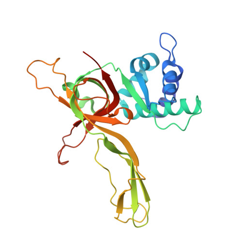
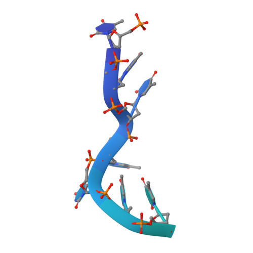
A conserved MCM single-stranded DNA binding element is essential for replication initiation.

Froelich, C.A.Kang, S.Epling, L.B.Bell, S.P.Enemark, E.J.

(2014) Elife 3: e01993-e01993

  • DOI: https://doi.org/10.7554/eLife.01993
  • Primary Citation of Related Structures:  
    4POF, 4POG

  • PubMed Abstract: 

    The ring-shaped MCM helicase is essential to all phases of DNA replication. The complex loads at replication origins as an inactive double-hexamer encircling duplex DNA. Helicase activation converts this species to two active single hexamers that encircle single-stranded DNA (ssDNA). The molecular details of MCM DNA interactions during these events are unknown. We determined the crystal structure of the Pyrococcus furiosus MCM N-terminal domain hexamer bound to ssDNA and define a conserved MCM-ssDNA binding motif (MSSB). Intriguingly, ssDNA binds the MCM ring interior perpendicular to the central channel with defined polarity. In eukaryotes, the MSSB is conserved in several Mcm2-7 subunits, and MSSB mutant combinations in S. cerevisiae Mcm2-7 are not viable. Mutant Mcm2-7 complexes assemble and are recruited to replication origins, but are defective in helicase loading and activation. Our findings identify an important MCM-ssDNA interaction and suggest it functions during helicase activation to select the strand for translocation. DOI: http://dx.doi.org/10.7554/eLife.01993.001.


  • Organizational Affiliation

    Department of Structural Biology, St Jude Children's Research Hospital, Memphis, United States.


Macromolecules

Find similar proteins by:  (by identity cutoff)  |  3D Structure
Entity ID: 1
MoleculeChains Sequence LengthOrganismDetailsImage
Cell division control protein 21
A, B, C, D, E
A, B, C, D, E, F, G, H, I, J, K, L
257Pyrococcus furiosus DSM 3638Mutation(s): 0 
Gene Names: Cdc21PF0482
UniProt
Find proteins for Q8U3I4 (Pyrococcus furiosus (strain ATCC 43587 / DSM 3638 / JCM 8422 / Vc1))
Explore Q8U3I4 
Go to UniProtKB:  Q8U3I4
Entity Groups  
Sequence Clusters30% Identity50% Identity70% Identity90% Identity95% Identity100% Identity
UniProt GroupQ8U3I4
Sequence Annotations
Expand
  • Reference Sequence
Find similar nucleic acids by:  (by identity cutoff)  |  3D Structure
Entity ID: 2
MoleculeChains LengthOrganismImage
30-mer oligo(dT)M [auth X],
N [auth V],
O [auth Y],
P [auth Z]
30N/A
Sequence Annotations
Expand
  • Reference Sequence
Small Molecules
Ligands 1 Unique
IDChains Name / Formula / InChI Key2D Diagram3D Interactions
ZN
Query on ZN

Download Ideal Coordinates CCD File 
AA [auth K]
BA [auth L]
Q [auth A]
R [auth B]
S [auth C]
AA [auth K],
BA [auth L],
Q [auth A],
R [auth B],
S [auth C],
T [auth D],
U [auth E],
V [auth F],
W [auth G],
X [auth H],
Y [auth I],
Z [auth J]
ZINC ION
Zn
PTFCDOFLOPIGGS-UHFFFAOYSA-N
Experimental Data & Validation

Experimental Data

  • Method: X-RAY DIFFRACTION
  • Resolution: 3.20 Å
  • R-Value Free: 0.294 
  • R-Value Work: 0.257 
  • R-Value Observed: 0.259 
  • Space Group: P 1 21 1
Unit Cell:
Length ( Å )Angle ( ˚ )
a = 94.277α = 90
b = 113.398β = 101.35
c = 196.855γ = 90
Software Package:
Software NamePurpose
SERGUIdata collection
PHASERphasing
REFMACrefinement
HKL-2000data reduction
HKL-2000data scaling

Structure Validation

View Full Validation Report


Entry History 

Deposition Data

Revision History  (Full details and data files)

  • Version 1.0: 2014-04-09
    Type: Initial release
  • Version 1.1: 2014-12-24
    Changes: Database references
  • Version 1.2: 2017-11-22
    Changes: Refinement description
  • Version 1.3: 2023-09-20
    Changes: Data collection, Database references, Derived calculations, Refinement description