4PN1

Structure of S. pombe Pct1 RNA triphosphatase in complex with the Spt5 CTD


Experimental Data Snapshot

  • Method: X-RAY DIFFRACTION
  • Resolution: 2.80 Å
  • R-Value Free: 0.248 
  • R-Value Work: 0.191 
  • R-Value Observed: 0.194 

wwPDB Validation   3D Report Full Report


This is version 1.5 of the entry. See complete history


Literature

Fission yeast RNA triphosphatase reads an Spt5 CTD code.

Doamekpor, S.K.Schwer, B.Sanchez, A.M.Shuman, S.Lima, C.D.

(2015) RNA 21: 113-123

  • DOI: https://doi.org/10.1261/rna.048181.114
  • Primary Citation of Related Structures:  
    4PN0, 4PN1

  • PubMed Abstract: 

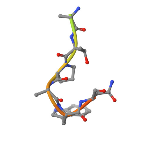
    mRNA capping enzymes are directed to nascent RNA polymerase II (Pol2) transcripts via interactions with the carboxy-terminal domains (CTDs) of Pol2 and transcription elongation factor Spt5. Fission yeast RNA triphosphatase binds to the Spt5 CTD, comprising a tandem repeat of nonapeptide motif TPAWNSGSK. Here we report the crystal structure of a Pct1·Spt5-CTD complex, which revealed two CTD docking sites on the Pct1 homodimer that engage TPAWN segments of the motif. Each Spt5 CTD interface, composed of elements from both subunits of the homodimer, is dominated by van der Waals contacts from Pct1 to the tryptophan of the CTD. The bound CTD adopts a distinctive conformation in which the peptide backbone makes a tight U-turn so that the proline stacks over the tryptophan. We show that Pct1 binding to Spt5 CTD is antagonized by threonine phosphorylation. Our results fortify an emerging concept of an "Spt5 CTD code" in which (i) the Spt5 CTD is structurally plastic and can adopt different conformations that are templated by particular cellular Spt5 CTD receptor proteins; and (ii) threonine phosphorylation of the Spt5 CTD repeat inscribes a binary on-off switch that is read by diverse CTD receptors, each in its own distinctive manner.


  • Organizational Affiliation

    Structural Biology Program, Sloan-Kettering Institute, New York, New York 10065, USA.


Macromolecules
Find similar proteins by:  (by identity cutoff)  |  3D Structure
Entity ID: 1
MoleculeChains Sequence LengthOrganismDetailsImage
mRNA-capping enzyme subunit beta
A, B, C, D
304Schizosaccharomyces pombeMutation(s): 0 
Gene Names: pct1
EC: 3.1.3.33
UniProt
Find proteins for Q9P6Q6 (Schizosaccharomyces pombe (strain 972 / ATCC 24843))
Explore Q9P6Q6 
Go to UniProtKB:  Q9P6Q6
Entity Groups  
Sequence Clusters30% Identity50% Identity70% Identity90% Identity95% Identity100% Identity
UniProt GroupQ9P6Q6
Sequence Annotations
Expand
  • Reference Sequence

Find similar proteins by:  Sequence   |   3D Structure  

Entity ID: 2
MoleculeChains Sequence LengthOrganismDetailsImage
Synthetic peptide
E, F, G, H
18N/AMutation(s): 0 
Entity Groups  
Sequence Clusters30% Identity50% Identity70% Identity90% Identity95% Identity100% Identity
Sequence Annotations
Expand
  • Reference Sequence
Experimental Data & Validation

Experimental Data

  • Method: X-RAY DIFFRACTION
  • Resolution: 2.80 Å
  • R-Value Free: 0.248 
  • R-Value Work: 0.191 
  • R-Value Observed: 0.194 
  • Space Group: P 21 21 2
Unit Cell:
Length ( Å )Angle ( ˚ )
a = 95.603α = 90
b = 251.542β = 90
c = 75.397γ = 90
Software Package:
Software NamePurpose
PHENIXrefinement

Structure Validation

View Full Validation Report


Entry History & Funding Information

Deposition Data


Funding OrganizationLocationGrant Number
National Institutes of Health/National Institute of General Medical Sciences (NIH/NIGMS)United StatesGM061906

Revision History  (Full details and data files)

  • Version 1.0: 2014-11-12
    Type: Initial release
  • Version 1.1: 2014-12-10
    Changes: Database references
  • Version 1.2: 2014-12-31
    Changes: Database references
  • Version 1.3: 2017-09-27
    Changes: Author supporting evidence, Database references, Derived calculations, Other, Source and taxonomy
  • Version 1.4: 2019-12-25
    Changes: Author supporting evidence
  • Version 1.5: 2023-09-27
    Changes: Data collection, Database references, Refinement description