4PL3

Crystal structure of murine IRE1 in complex with MKC9989 inhibitor


Experimental Data Snapshot

  • Method: X-RAY DIFFRACTION
  • Resolution: 2.90 Å
  • R-Value Free: 0.234 
  • R-Value Work: 0.201 
  • R-Value Observed: 0.202 

wwPDB Validation   3D Report Full Report


Ligand Structure Quality Assessment 


This is version 1.4 of the entry. See complete history


Literature

Structure and mechanism of action of the hydroxy-aryl-aldehyde class of IRE1 endoribonuclease inhibitors.

Sanches, M.Duffy, N.M.Talukdar, M.Thevakumaran, N.Chiovitti, D.Canny, M.D.Lee, K.Kurinov, I.Uehling, D.Al-Awar, R.Poda, G.Prakesch, M.Wilson, B.Tam, V.Schweitzer, C.Toro, A.Lucas, J.L.Vuga, D.Lehmann, L.Durocher, D.Zeng, Q.Patterson, J.B.Sicheri, F.

(2014) Nat Commun 5: 4202-4202

  • DOI: https://doi.org/10.1038/ncomms5202
  • Primary Citation of Related Structures:  
    4PL3, 4PL4, 4PL5

  • PubMed Abstract: 

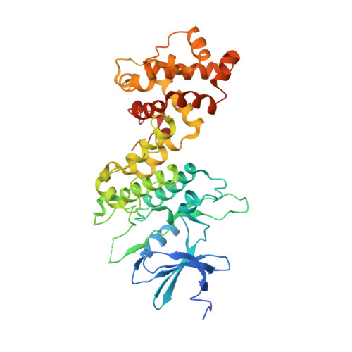
    Endoplasmic reticulum (ER) stress activates the unfolded protein response and its dysfunction is linked to multiple diseases. The stress transducer IRE1α is a transmembrane kinase endoribonuclease (RNase) that cleaves mRNA substrates to re-establish ER homeostasis. Aromatic ring systems containing hydroxy-aldehyde moieties, termed hydroxy-aryl-aldehydes (HAA), selectively inhibit IRE1α RNase and thus represent a novel chemical series for therapeutic development. We solved crystal structures of murine IRE1α in complex with three HAA inhibitors. HAA inhibitors engage a shallow pocket at the RNase-active site through pi-stacking interactions with His910 and Phe889, an essential Schiff base with Lys907 and a hydrogen bond with Tyr892. Structure-activity studies and mutational analysis of contact residues define the optimal chemical space of inhibitors and validate the inhibitor-binding site. These studies lay the foundation for understanding both the biochemical and cellular functions of IRE1α using small molecule inhibitors and suggest new avenues for inhibitor design.


  • Organizational Affiliation

    1] Centre for Systems Biology, Lunenfeld-Tanenbaum Research Institute, Mount Sinai Hospital, Toronto, Ontario, Canada M5G 1X5 [2].


Macromolecules
Find similar proteins by:  (by identity cutoff)  |  3D Structure
Entity ID: 1
MoleculeChains Sequence LengthOrganismDetailsImage
Serine/threonine-protein kinase/endoribonuclease IRE1
A, B
435Mus musculusMutation(s): 1 
Gene Names: Ern1Ire1
EC: 2.7.11.1 (PDB Primary Data), 3.1.26 (PDB Primary Data)
UniProt
Find proteins for Q9EQY0 (Mus musculus)
Explore Q9EQY0 
Go to UniProtKB:  Q9EQY0
Entity Groups  
Sequence Clusters30% Identity50% Identity70% Identity90% Identity95% Identity100% Identity
UniProt GroupQ9EQY0
Sequence Annotations
Expand
  • Reference Sequence
Experimental Data & Validation

Experimental Data

  • Method: X-RAY DIFFRACTION
  • Resolution: 2.90 Å
  • R-Value Free: 0.234 
  • R-Value Work: 0.201 
  • R-Value Observed: 0.202 
  • Space Group: C 1 2 1
Unit Cell:
Length ( Å )Angle ( ˚ )
a = 246.08α = 90
b = 90.5β = 91.88
c = 72.13γ = 90
Software Package:
Software NamePurpose
PHENIXrefinement
XDSdata reduction
XSCALEdata scaling
PDB_EXTRACTdata extraction
PHASERphasing

Structure Validation

View Full Validation Report


Ligand Structure Quality Assessment 


Entry History & Funding Information

Deposition Data


Funding OrganizationLocationGrant Number
Canadian Institutes of Health Research (CIHR)CanadaMOP 84370
Multiple Myeloma Research Foundation Biotech Investment AwardUnited States--
Canadian Cancer SocietyCanada--

Revision History  (Full details and data files)

  • Version 1.0: 2014-09-03
    Type: Initial release
  • Version 1.1: 2014-09-10
    Changes: Database references
  • Version 1.2: 2017-11-22
    Changes: Advisory, Derived calculations, Other, Refinement description, Source and taxonomy
  • Version 1.3: 2020-01-08
    Changes: Author supporting evidence
  • Version 1.4: 2023-09-27
    Changes: Data collection, Database references, Derived calculations, Refinement description