Structural Insight into Host Recognition by Aggregative Adherence Fimbriae of Enteroaggregative Escherichia coli.
Berry, A.A., Yang, Y., Pakharukova, N., Garnett, J.A., Lee, W.C., Cota, E., Marchant, J., Roy, S., Tuittila, M., Liu, B., Inman, K.G., Ruiz-Perez, F., Mandomando, I., Nataro, J.P., Zavialov, A.V., Matthews, S.(2014) PLoS Pathog 10: e1004404-e1004404
- PubMed: 25232738
- DOI: https://doi.org/10.1371/journal.ppat.1004404
- Primary Citation of Related Structures:
2MPV, 4OR1, 4PH8, 4PHX - PubMed Abstract:
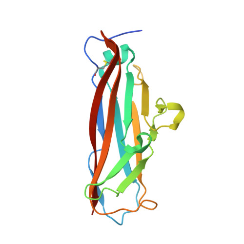
Enteroaggregative Escherichia coli (EAEC) is a leading cause of acute and persistent diarrhea worldwide. A recently emerged Shiga-toxin-producing strain of EAEC resulted in significant mortality and morbidity due to progressive development of hemolytic-uremic syndrome. The attachment of EAEC to the human intestinal mucosa is mediated by aggregative adherence fimbria (AAF). Using X-ray crystallography and NMR structures, we present new atomic resolution insight into the structure of AAF variant I from the strain that caused the deadly outbreak in Germany in 2011, and AAF variant II from archetype strain 042, and propose a mechanism for AAF-mediated adhesion and biofilm formation. Our work shows that major subunits of AAF assemble into linear polymers by donor strand complementation where a single minor subunit is inserted at the tip of the polymer by accepting the donor strand from the terminal major subunit. Whereas the minor subunits of AAF have a distinct conserved structure, AAF major subunits display large structural differences, affecting the overall pilus architecture. These structures suggest a mechanism for AAF-mediated adhesion and biofilm formation. Binding experiments using wild type and mutant subunits (NMR and SPR) and bacteria (ELISA) revealed that despite the structural differences AAF recognize a common receptor, fibronectin, by employing clusters of basic residues at the junction between subunits in the pilus. We show that AAF-fibronectin attachment is based primarily on electrostatic interactions, a mechanism not reported previously for bacterial adhesion to biotic surfaces.
Organizational Affiliation:
Center for Vaccine Development, Department of Pediatrics, University of Maryland School of Medicine, Baltimore, Maryland, United States of America.