4PGA

GLUTAMINASE-ASPARAGINASE FROM PSEUDOMONAS 7A


Experimental Data Snapshot

  • Method: X-RAY DIFFRACTION
  • Resolution: 1.70 Å
  • R-Value Free: 0.254 
  • R-Value Work: 0.199 
  • R-Value Observed: 0.199 

wwPDB Validation   3D Report Full Report


This is version 1.3 of the entry. See complete history


Literature

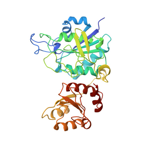
Ion binding induces closed conformation in Pseudomonas 7A glutaminase-asparaginase (PGA): crystal structure of the PGA-SO4(2-)-NH4+ complex at 1.7 A resolution.

Jakob, C.G.Lewinski, K.LaCount, M.W.Roberts, J.Lebioda, L.

(1997) Biochemistry 36: 923-931

  • DOI: https://doi.org/10.1021/bi961979x
  • Primary Citation of Related Structures:  
    4PGA

  • PubMed Abstract: 

    Pseudomonas 7A glutaminase-asparaginase (PGA) catalyzes the hydrolysis of D- and L-isomers of glutamine and asparagine. X-ray quality type-1 crystals of PGA have been obtained from 2.0 M ammonium sulfate. The space group is C222(1) with unit-cell dimensions a = 78.62, b = 135.80, and c = 137.88 A. The tetrameric molecule is located on a crystallographic 2-fold axis, and two subunits form the asymmetric portion of the unit cell. The structure was solved by the molecular replacement method and refined at 1.7 A resolution to an R = 19.9% with a good geometry of the model, G = 0.05. The resultant electron density maps enabled us to resolve individual constituent atoms of most residues and introduce minor revisions to the amino acid sequence. The catalytic loop, Thr20-Gly40, is in the closed conformation with excellent electron density in both subunits. A sulfate ion and an ammonium ion are bound in the substrate binding site and interect with the loop. This interaction appears to be responsible for the observed closed conformation. New arguments supporting Thr20 as the catalytic nucleophile in the asparaginase activity are proposed.


  • Organizational Affiliation

    Department of Chemistry and Biochemistry, University of South Carolina, Columbia 29208, USA.


Macromolecules
Find similar proteins by:  (by identity cutoff)  |  3D Structure
Entity ID: 1
MoleculeChains Sequence LengthOrganismDetailsImage
GLUTAMINASE-ASPARAGINASE
A, B
337Pseudomonas sp. 7AMutation(s): 0 
EC: 3.5.1.38
UniProt
Find proteins for P10182 (Pseudomonas sp. (strain ATCC 29598 / 7A))
Explore P10182 
Go to UniProtKB:  P10182
Entity Groups  
Sequence Clusters30% Identity50% Identity70% Identity90% Identity95% Identity100% Identity
UniProt GroupP10182
Sequence Annotations
Expand
  • Reference Sequence
Experimental Data & Validation

Experimental Data

  • Method: X-RAY DIFFRACTION
  • Resolution: 1.70 Å
  • R-Value Free: 0.254 
  • R-Value Work: 0.199 
  • R-Value Observed: 0.199 
  • Space Group: C 2 2 21
Unit Cell:
Length ( Å )Angle ( ˚ )
a = 78.62α = 90
b = 135.8β = 90
c = 137.88γ = 90
Software Package:
Software NamePurpose
X-PLORmodel building
X-PLORrefinement
bioteXdata reduction
bioteXdata scaling
X-PLORphasing

Structure Validation

View Full Validation Report


Entry History 

Deposition Data

Revision History  (Full details and data files)

  • Version 1.0: 1997-07-23
    Type: Initial release
  • Version 1.1: 2008-03-25
    Changes: Version format compliance
  • Version 1.2: 2011-07-13
    Changes: Derived calculations, Version format compliance
  • Version 1.3: 2023-08-09
    Changes: Database references, Derived calculations, Other, Refinement description