


Crystal Structure of Cobyrinic Acid a,c-diamide synthase from Mycobacterium smegmatis
Davies, D.R., Fairman, J., Abendroth, J.To be published.
Experimental Data Snapshot
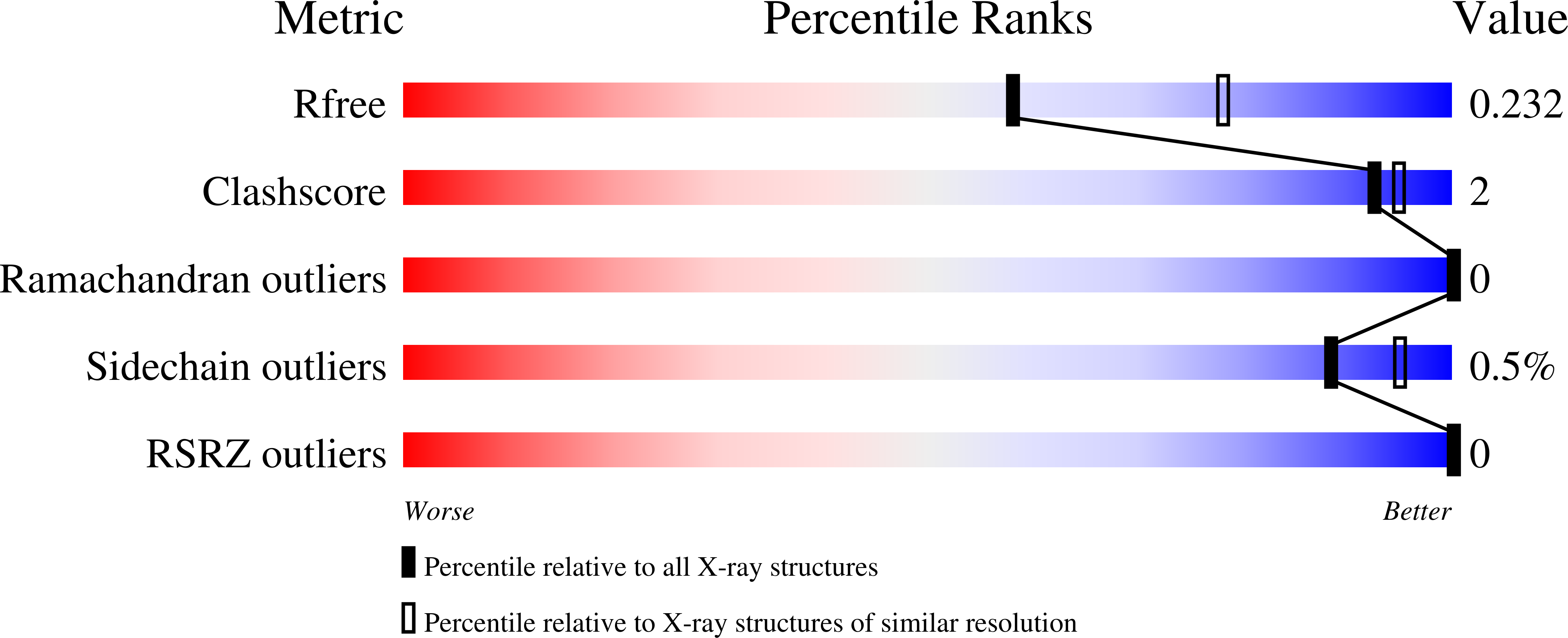
wwPDB Validation 3D Report Full Report
Entity ID: 1 | |||||
|---|---|---|---|---|---|
| Molecule | Chains | Sequence Length | Organism | Details | Image |
| Cobyrinic Acid a,c-diamide synthase | 273 | Mycolicibacterium smegmatis MC2 155 | Mutation(s): 0 Gene Names: MSMEG_1927 | 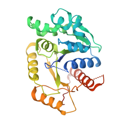 | |
UniProt | |||||
Find proteins for A0QTQ5 (Mycolicibacterium smegmatis (strain ATCC 700084 / mc(2)155)) Explore A0QTQ5 Go to UniProtKB: A0QTQ5 | |||||
Entity Groups | |||||
| Sequence Clusters | 30% Identity50% Identity70% Identity90% Identity95% Identity100% Identity | ||||
| UniProt Group | A0QTQ5 | ||||
Sequence AnnotationsExpand | |||||
| |||||
| Length ( Å ) | Angle ( ˚ ) |
|---|---|
| a = 88.81 | α = 90 |
| b = 88.81 | β = 90 |
| c = 122.31 | γ = 90 |
| Software Name | Purpose |
|---|---|
| BALBES | phasing |
| PHENIX | refinement |
RCSB PDB (citation) is hosted by
RCSB PDB is a member of the

