


Crystal structure of M. tuberculosis in complex with BTO
Neres, J., Cole, S.To be published.
Experimental Data Snapshot
Entity ID: 1 | |||||
|---|---|---|---|---|---|
| Molecule | Chains | Sequence Length | Organism | Details | Image |
| Probable decaprenylphosphoryl-beta-D-ribose oxidase | 480 | Mycobacterium tuberculosis H37Rv | Mutation(s): 0 Gene Names: dprE1, Rv3790 EC: 1 | 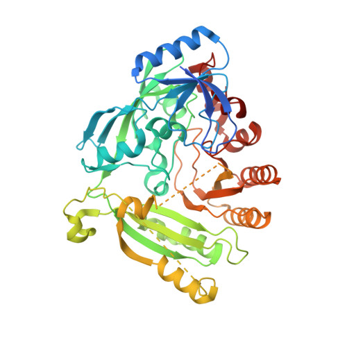 | |
UniProt | |||||
Find proteins for P9WJF1 (Mycobacterium tuberculosis (strain ATCC 25618 / H37Rv)) Explore P9WJF1 Go to UniProtKB: P9WJF1 | |||||
Entity Groups | |||||
| Sequence Clusters | 30% Identity50% Identity70% Identity90% Identity95% Identity100% Identity | ||||
| UniProt Group | P9WJF1 | ||||
Sequence AnnotationsExpand | |||||
| |||||
| Ligands 2 Unique | |||||
|---|---|---|---|---|---|
| ID | Chains | Name / Formula / InChI Key | 2D Diagram | 3D Interactions | |
| FAD Query on FAD | C [auth A], E [auth B] | FLAVIN-ADENINE DINUCLEOTIDE C27 H33 N9 O15 P2 VWWQXMAJTJZDQX-UYBVJOGSSA-N |  | ||
| N77 Query on N77 | D [auth A], F [auth B] | 7-(hydroxyamino)-N-(pyridin-3-ylmethyl)-5-(trifluoromethyl)-1,3-benzothiazole-2-carboxamide 3-oxide C15 H11 F3 N4 O3 S GENRUIYZXCGKPM-UHFFFAOYSA-N |  | ||
| Length ( Å ) | Angle ( ˚ ) |
|---|---|
| a = 72.856 | α = 90 |
| b = 82.013 | β = 100.77 |
| c = 81.148 | γ = 90 |
| Software Name | Purpose |
|---|---|
| REFMAC | refinement |
| XDS | data scaling |
| PHASER | phasing |
| Funding Organization | Location | Grant Number |
|---|---|---|
| European Commission | Switzerland | MM4TB - 260872 |
RCSB PDB (citation) is hosted by
RCSB PDB is a member of the

