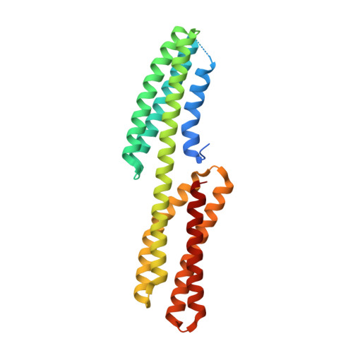
4P9T

Structure of the free form of the N-terminal VH1 domain of monomeric alpha-catenin


Experimental Data Snapshot

  • Method: X-RAY DIFFRACTION
  • Resolution: 2.50 Å
  • R-Value Free: 0.234 
  • R-Value Work: 0.215 
  • R-Value Observed: 0.216 

wwPDB Validation   3D Report Full Report


This is version 1.4 of the entry. See complete history


Literature

Structure of the free form of the N-terminal VH1 domain of monomeric alpha-catenin.

Shibahara, T.Hirano, Y.Hakoshima, T.

(2015) FEBS Lett 589: 1754-1760

  • DOI: https://doi.org/10.1016/j.febslet.2015.05.053
  • Primary Citation of Related Structures:  
    4P9T

  • PubMed Abstract: 

    The N-terminal vinculin-homology 1 (VH1) domain of α-catenin facilitates two exclusive forms, a monomeric form directly bound to β-catenin for linking E-cadherin to F-actin or a homodimer for the inhibition of β-catenin binding. Competition of these two forms is affected by ∼80 N-terminal residues, whose structure is poorly understood. We have determined the structure of the monomeric free form of the αN-catenin VH1 domain and revealed that the N-terminal residues form α1 and α2 helices to complete formation of the N-terminal four-helix bundle. Dynamic conformational changes of these two helices control formation of the β-catenin-bound monomer or unbound homodimer.


  • Organizational Affiliation

    Structural Biology Laboratory, Nara Institute of Science and Technology, 8916-5 Takayama, Ikoma, Nara 630-0192, Japan.


Macromolecules
Find similar proteins by:  (by identity cutoff)  |  3D Structure
Entity ID: 1
MoleculeChains Sequence LengthOrganismDetailsImage
Catenin alpha-2
A, B, C, D
263Mus musculusMutation(s): 0 
Gene Names: Ctnna2Catna2
UniProt
Find proteins for Q61301 (Mus musculus)
Explore Q61301 
Go to UniProtKB:  Q61301
Entity Groups  
Sequence Clusters30% Identity50% Identity70% Identity90% Identity95% Identity100% Identity
UniProt GroupQ61301
Sequence Annotations
Expand
  • Reference Sequence
Small Molecules
Ligands 3 Unique
IDChains Name / Formula / InChI Key2D Diagram3D Interactions
IOD
Query on IOD

Download Ideal Coordinates CCD File 
E [auth A]
EA [auth D]
F [auth A]
FA [auth D]
G [auth A]
E [auth A],
EA [auth D],
F [auth A],
FA [auth D],
G [auth A],
GA [auth D],
H [auth A],
HA [auth D],
I [auth A],
IA [auth D],
N [auth B],
O [auth B],
P [auth B],
Q [auth B],
R [auth B],
V [auth C],
W [auth C],
X [auth C],
Y [auth C],
Z [auth C]
IODIDE ION
I
XMBWDFGMSWQBCA-UHFFFAOYSA-M
PEG
Query on PEG

Download Ideal Coordinates CCD File 
DA [auth C],
M [auth A]
DI(HYDROXYETHYL)ETHER
C4 H10 O3
MTHSVFCYNBDYFN-UHFFFAOYSA-N
EDO
Query on EDO

Download Ideal Coordinates CCD File 
AA [auth C]
BA [auth C]
CA [auth C]
J [auth A]
JA [auth D]
AA [auth C],
BA [auth C],
CA [auth C],
J [auth A],
JA [auth D],
K [auth A],
KA [auth D],
L [auth A],
LA [auth D],
S [auth B],
T [auth B],
U [auth B]
1,2-ETHANEDIOL
C2 H6 O2
LYCAIKOWRPUZTN-UHFFFAOYSA-N
Experimental Data & Validation

Experimental Data

  • Method: X-RAY DIFFRACTION
  • Resolution: 2.50 Å
  • R-Value Free: 0.234 
  • R-Value Work: 0.215 
  • R-Value Observed: 0.216 
  • Space Group: P 31
Unit Cell:
Length ( Å )Angle ( ˚ )
a = 68.777α = 90
b = 68.777β = 90
c = 207.965γ = 120
Software Package:
Software NamePurpose
PHENIXrefinement
HKL-2000data scaling
MrBUMPphasing
Cootmodel building

Structure Validation

View Full Validation Report


Entry History 

Deposition Data

Revision History  (Full details and data files)

  • Version 1.0: 2015-04-29
    Type: Initial release
  • Version 1.1: 2015-06-10
    Changes: Database references
  • Version 1.2: 2015-07-15
    Changes: Database references
  • Version 1.3: 2017-11-22
    Changes: Data collection, Database references, Derived calculations, Other, Refinement description, Source and taxonomy, Structure summary
  • Version 1.4: 2023-09-27
    Changes: Data collection, Database references, Refinement description