4P8S

Crystal structure of Nogo-receptor-2


Experimental Data Snapshot

  • Method: X-RAY DIFFRACTION
  • Resolution: 1.80 Å
  • R-Value Free: 0.205 
  • R-Value Work: 0.171 
  • R-Value Observed: 0.173 

wwPDB Validation   3D Report Full Report


Ligand Structure Quality Assessment 


This is version 2.1 of the entry. See complete history


Literature

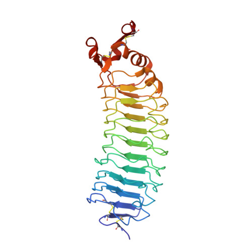
Crystal structure of the Nogo-receptor-2.

Semavina, M.Saha, N.Kolev, M.V.Goldgur, Y.Giger, R.J.Himanen, J.P.Nikolov, D.B.

(2011) Protein Sci : 684-689

  • DOI: https://doi.org/10.1002/pro.597
  • Primary Citation of Related Structures:  
    4P8S, 4P91

  • PubMed Abstract: 

    The inhibition of axon regeneration upon mechanical injury is dependent on interactions between Nogo receptors (NgRs) and their myelin-derived ligands. NgRs are composed of a leucine-rich repeat (LRR) region, thought to be structurally similar among the different isoforms of the receptor, and a divergent "stalk" region. It has been shown by others that the LRR and stalk regions of NgR1 and NgR2 have distinct roles in conferring binding affinity to the myelin associated glycoprotein (MAG) in vivo. Here, we show that purified recombinant full length NgR1 and NgR2 maintain significantly higher binding affinity for purified MAG as compared to the isolated LRR region of either NgR1 or NgR2. We also present the crystal structure of the LRR and part of the stalk regions of NgR2 and compare it to the previously reported NgR1 structure with respect to the distinct signaling properties of the two receptor isoforms.


  • Organizational Affiliation

    Structural Biology Program, Memorial Sloan Kettering Cancer Center, 1275 York Avenue, New York 10065, USA.


Macromolecules
Find similar proteins by:  (by identity cutoff)  |  3D Structure
Entity ID: 1
MoleculeChains Sequence LengthOrganismDetailsImage
Reticulon-4 receptor-like 2282Rattus norvegicusMutation(s): 0 
Gene Names: Rtn4rl2Ngrh1
UniProt
Find proteins for Q80WD1 (Rattus norvegicus)
Explore Q80WD1 
Go to UniProtKB:  Q80WD1
Entity Groups  
Sequence Clusters30% Identity50% Identity70% Identity90% Identity95% Identity100% Identity
UniProt GroupQ80WD1
Sequence Annotations
Expand
  • Reference Sequence
Oligosaccharides

Help

Entity ID: 2
MoleculeChains Length2D Diagram Glycosylation3D Interactions
2-acetamido-2-deoxy-alpha-D-glucopyranose-(1-4)-2-acetamido-2-deoxy-beta-D-glucopyranose
B
2N-Glycosylation
Glycosylation Resources
GlyTouCan:  G07375KG
GlyCosmos:  G07375KG
GlyGen:  G07375KG
Entity ID: 3
MoleculeChains Length2D Diagram Glycosylation3D Interactions
2-acetamido-2-deoxy-beta-D-glucopyranose-(1-4)-2-acetamido-2-deoxy-beta-D-glucopyranose
C
2N-Glycosylation
Glycosylation Resources
GlyTouCan:  G42666HT
GlyCosmos:  G42666HT
GlyGen:  G42666HT
Small Molecules
Ligands 1 Unique
IDChains Name / Formula / InChI Key2D Diagram3D Interactions
NAG
Query on NAG

Download Ideal Coordinates CCD File 
D [auth A]2-acetamido-2-deoxy-beta-D-glucopyranose
C8 H15 N O6
OVRNDRQMDRJTHS-FMDGEEDCSA-N
Experimental Data & Validation

Experimental Data

  • Method: X-RAY DIFFRACTION
  • Resolution: 1.80 Å
  • R-Value Free: 0.205 
  • R-Value Work: 0.171 
  • R-Value Observed: 0.173 
  • Space Group: P 21 21 21
Unit Cell:
Length ( Å )Angle ( ˚ )
a = 56.822α = 90
b = 56.517β = 90
c = 128.96γ = 90
Software Package:
Software NamePurpose
REFMACrefinement

Structure Validation

View Full Validation Report


Ligand Structure Quality Assessment 


Entry History 

Deposition Data

Revision History  (Full details and data files)

  • Version 1.0: 2014-04-09
    Type: Initial release
  • Version 1.1: 2015-02-11
    Changes: Non-polymer description
  • Version 2.0: 2020-07-29
    Type: Remediation
    Reason: Carbohydrate remediation
    Changes: Advisory, Atomic model, Data collection, Derived calculations, Source and taxonomy, Structure summary
  • Version 2.1: 2023-12-27
    Changes: Data collection, Database references, Structure summary