4P58

Crystal structure of mouse comt bound to an inhibitor


Experimental Data Snapshot

  • Method: X-RAY DIFFRACTION
  • Resolution: 2.06 Å
  • R-Value Free: 0.199 
  • R-Value Work: 0.145 
  • R-Value Observed: 0.147 

wwPDB Validation   3D Report Full Report


Ligand Structure Quality Assessment 


This is version 1.2 of the entry. See complete history


Literature

A fragment-based approach to identifying S-adenosyl-l-methionine -competitive inhibitors of catechol O-methyl transferase (COMT).

Lanier, M.Ambrus, G.Cole, D.C.Davenport, R.Ellery, J.Fosbeary, R.Jennings, A.J.Kadotani, A.Kamada, Y.Kamran, R.Matsumoto, S.Mizukami, A.Okubo, S.Okada, K.Saikatendu, K.Walsh, L.Wu, H.Hixon, M.S.

(2014) J Med Chem 57: 5459-5463

  • DOI: https://doi.org/10.1021/jm500475k
  • Primary Citation of Related Structures:  
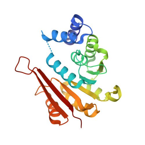
    4P58

  • PubMed Abstract: 

    Catechol O-methyl transferase belongs to the diverse family of S-adenosyl-l-methionine transferases. It is a target involved in the treatment of Parkinson's disease. Here we present a fragment-based screening approach to discover noncatechol derived COMT inhibitors which bind at the SAM binding pocket. We describe the identification and characterization of a series of highly ligand efficient SAM competitive bisaryl fragments (LE = 0.33-0.58). We also present the first SAM-competitive small-molecule COMT co-complex crystal structure.


  • Organizational Affiliation

    Medicinal Chemistry, ‡Structural Biology, §Discovery Biology, ∥Analytical Chemistry, Takeda California Inc. , 10410 Science Center Drive San Diego California 92121, United States.


Macromolecules
Find similar proteins by:  (by identity cutoff)  |  3D Structure
Entity ID: 1
MoleculeChains Sequence LengthOrganismDetailsImage
Catechol O-methyltransferase212Mus musculusMutation(s): 0 
Gene Names: ComtComt1
EC: 2.1.1.6
UniProt & NIH Common Fund Data Resources
Find proteins for O88587 (Mus musculus)
Explore O88587 
Go to UniProtKB:  O88587
IMPC:  MGI:88470
Entity Groups  
Sequence Clusters30% Identity50% Identity70% Identity90% Identity95% Identity100% Identity
UniProt GroupO88587
Sequence Annotations
Expand
  • Reference Sequence
Small Molecules
Ligands 1 Unique
IDChains Name / Formula / InChI Key2D Diagram3D Interactions
2F6
Query on 2F6

Download Ideal Coordinates CCD File 
B [auth A]1',3'-dimethyl-1H,1'H-3,4'-bipyrazole
C8 H10 N4
LNJKDWJFZASOPC-UHFFFAOYSA-N
Binding Affinity Annotations 
IDSourceBinding Affinity
2F6 BindingDB:  4P58 Ki: min: 6.30e+4, max: 1.58e+5 (nM) from 2 assay(s)
Binding MOAD:  4P58 Kd: 6.30e+4 (nM) from 1 assay(s)
Experimental Data & Validation

Experimental Data

  • Method: X-RAY DIFFRACTION
  • Resolution: 2.06 Å
  • R-Value Free: 0.199 
  • R-Value Work: 0.145 
  • R-Value Observed: 0.147 
  • Space Group: I 41 2 2
Unit Cell:
Length ( Å )Angle ( ˚ )
a = 98.584α = 90
b = 98.584β = 90
c = 115.104γ = 90
Software Package:
Software NamePurpose
REFMACrefinement
PDB_EXTRACTdata extraction

Structure Validation

View Full Validation Report


Ligand Structure Quality Assessment 


Entry History 

Deposition Data

  • Released Date: 2014-06-25 
  • Deposition Author(s): Lanier, M.

Revision History  (Full details and data files)

  • Version 1.0: 2014-06-25
    Type: Initial release
  • Version 1.1: 2014-10-01
    Changes: Database references
  • Version 1.2: 2023-09-27
    Changes: Data collection, Database references, Derived calculations, Other, Refinement description, Source and taxonomy, Structure summary