4P42

Extended-Synaptotagmin 2, SMP - C2A - C2B Domains


Experimental Data Snapshot

  • Method: X-RAY DIFFRACTION
  • Resolution: 2.44 Å
  • R-Value Free: 0.256 
  • R-Value Work: 0.210 
  • R-Value Observed: 0.212 

wwPDB Validation   3D Report Full Report


Ligand Structure Quality Assessment 


This is version 2.0 of the entry. See complete history


Literature

Structure of a lipid-bound extended synaptotagmin indicates a role in lipid transfer.

Schauder, C.M.Wu, X.Saheki, Y.Narayanaswamy, P.Torta, F.Wenk, M.R.De Camilli, P.Reinisch, K.M.

(2014) Nature 510: 552-555

  • DOI: https://doi.org/10.1038/nature13269
  • Primary Citation of Related Structures:  
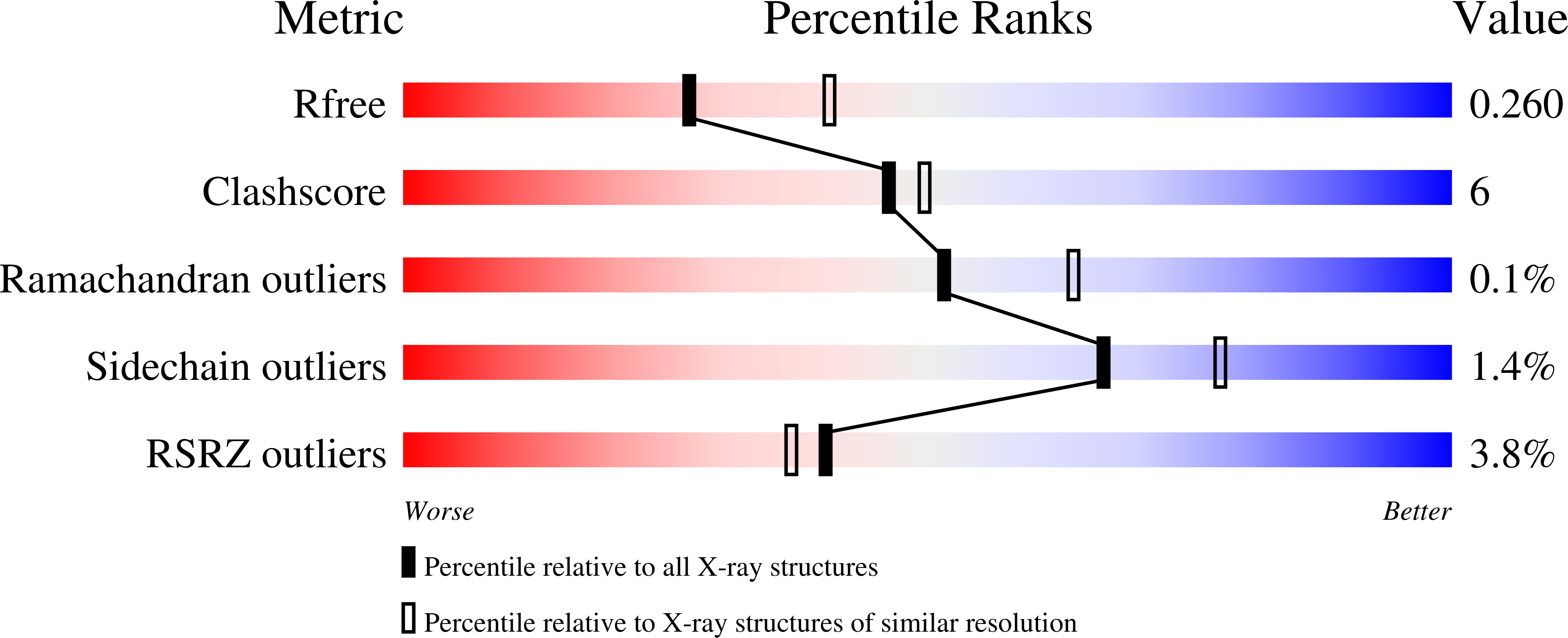
    4P42

  • PubMed Abstract: 

    Growing evidence suggests that close appositions between the endoplasmic reticulum (ER) and other membranes, including appositions with the plasma membrane (PM), mediate exchange of lipids between these bilayers. The mechanisms of such exchange, which allows lipid transfer independently of vesicular transport, remain poorly understood. The presence of a synaptotagmin-like mitochondrial-lipid-binding protein (SMP) domain, a proposed lipid-binding module, in several proteins localized at membrane contact sites has raised the possibility that such domains may be implicated in lipid transport. SMP-containing proteins include components of the ERMES complex, an ER–mitochondrial tether, and the extended synaptotagmins (known as tricalbins in yeast), which are ER–PM tethers. Here we present at 2.44 Å resolution the crystal structure of a fragment of human extended synaptotagmin 2 (E-SYT2), including an SMP domain and two adjacent C2 domains. The SMP domain has a β-barrel structure like protein modules in the tubular-lipid-binding (TULIP) superfamily. It dimerizes to form an approximately 90-Å-long cylinder traversed by a channel lined entirely with hydrophobic residues, with the two C2A–C2B fragments forming arched structures flexibly linked to the SMP domain. Importantly, structural analysis complemented by mass spectrometry revealed the presence of glycerophospholipids in the E-SYT2 SMP channel, indicating a direct role for E-SYTs in lipid transport. These findings provide strong evidence for a role of SMP-domain-containing proteins in the control of lipid transfer at membrane contact sites and have broad implications beyond the field of ER-to-PM appositions.


Macromolecules
Find similar proteins by:  (by identity cutoff)  |  3D Structure
Entity ID: 1
MoleculeChains Sequence LengthOrganismDetailsImage
Extended synaptotagmin-2
A, B
474Homo sapiensMutation(s): 0 
Gene Names: ESYT2FAM62BKIAA1228
Membrane Entity: Yes 
UniProt & NIH Common Fund Data Resources
Find proteins for A0FGR8 (Homo sapiens)
Explore A0FGR8 
Go to UniProtKB:  A0FGR8
PHAROS:  A0FGR8
GTEx:  ENSG00000117868 
Entity Groups  
Sequence Clusters30% Identity50% Identity70% Identity90% Identity95% Identity100% Identity
UniProt GroupA0FGR8
Sequence Annotations
Expand
  • Reference Sequence
Small Molecules
Ligands 2 Unique
IDChains Name / Formula / InChI Key2D Diagram3D Interactions
PEE
Query on PEE

Download Ideal Coordinates CCD File 
D [auth A],
F [auth B]
1,2-dioleoyl-sn-glycero-3-phosphoethanolamine
C41 H78 N O8 P
MWRBNPKJOOWZPW-NYVOMTAGSA-N
EGC
Query on EGC

Download Ideal Coordinates CCD File 
C [auth A],
E [auth B]
2-(2-{2-[2-(2-{2-[2-(2-{2-[4-(1,1,3,3-TETRAMETHYL-BUTYL)-PHENOXY]-ETHOXY}-ETHOXY)-ETHOXY]-ETHOXY}-ETHOXY)-ETHOXY]-ETHOX Y}-ETHOXY)-ETHANOL
C32 H58 O10
VJYAJQFKKLYARJ-UHFFFAOYSA-N
Experimental Data & Validation

Experimental Data

  • Method: X-RAY DIFFRACTION
  • Resolution: 2.44 Å
  • R-Value Free: 0.256 
  • R-Value Work: 0.210 
  • R-Value Observed: 0.212 
  • Space Group: P 21 21 21
Unit Cell:
Length ( Å )Angle ( ˚ )
a = 74.332α = 90
b = 88.382β = 90
c = 167.19γ = 90
Software Package:
Software NamePurpose
PHENIXrefinement

Structure Validation

View Full Validation Report


Ligand Structure Quality Assessment 


Entry History 

Deposition Data

Revision History  (Full details and data files)

  • Version 1.0: 2014-05-14
    Type: Initial release
  • Version 1.1: 2014-06-18
    Changes: Database references
  • Version 1.2: 2014-07-16
    Changes: Database references
  • Version 2.0: 2023-12-27
    Changes: Advisory, Atomic model, Data collection, Database references, Derived calculations, Non-polymer description, Other, Refinement description, Source and taxonomy, Structure summary