4P3Y

Crystal structure of Acinetobacter baumannii DsbA in complex with EF-Tu


Experimental Data Snapshot

  • Method: X-RAY DIFFRACTION
  • Resolution: 2.15 Å
  • R-Value Free: 0.215 
  • R-Value Work: 0.170 
  • R-Value Observed: 0.172 

wwPDB Validation   3D Report Full Report


Ligand Structure Quality Assessment 


This is version 2.1 of the entry. See complete history


Literature

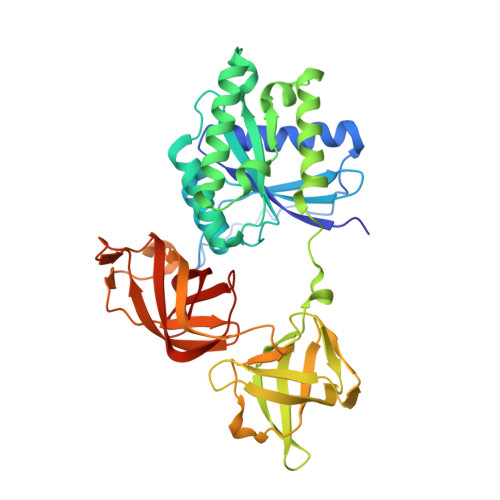
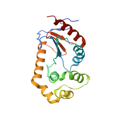
Structure of the Acinetobacter baumannii Dithiol Oxidase DsbA Bound to Elongation Factor EF-Tu Reveals a Novel Protein Interaction Site.

Premkumar, L.Kurth, F.Duprez, W.Grftehauge, M.K.King, G.J.Halili, M.A.Heras, B.Martin, J.L.

(2014) J Biol Chem 289: 19869-19880

  • DOI: https://doi.org/10.1074/jbc.M114.571737
  • Primary Citation of Related Structures:  
    4P3Y

  • PubMed Abstract: 

    The multidrug resistant bacterium Acinetobacter baumannii is a significant cause of nosocomial infection. Biofilm formation, that requires both disulfide bond forming and chaperone-usher pathways, is a major virulence trait in this bacterium. Our biochemical characterizations show that the periplasmic A. baumannii DsbA (AbDsbA) enzyme has an oxidizing redox potential and dithiol oxidase activity. We found an unexpected non-covalent interaction between AbDsbA and the highly conserved prokaryotic elongation factor, EF-Tu. EF-Tu is a cytoplasmic protein but has been localized extracellularly in many bacterial pathogens. The crystal structure of this complex revealed that the EF-Tu switch I region binds to the non-catalytic surface of AbDsbA. Although the physiological and pathological significance of a DsbA/EF-Tu association is unknown, peptides derived from the EF-Tu switch I region bound to AbDsbA with submicromolar affinity. We also identified a seven-residue DsbB-derived peptide that bound to AbDsbA with low micromolar affinity. Further characterization confirmed that the EF-Tu- and DsbB-derived peptides bind at two distinct sites. These data point to the possibility that the non-catalytic surface of DsbA is a potential substrate or regulatory protein interaction site. The two peptides identified in this work together with the newly characterized interaction site provide a novel starting point for inhibitor design targeting AbDsbA.


  • Organizational Affiliation

    From the Institute for Molecular Bioscience, Division of Chemistry and Structural Biology, University of Queensland, St. Lucia, Queensland 4067, Australia p.lakshmanane@uq.edu.au.


Macromolecules
Find similar proteins by:  (by identity cutoff)  |  3D Structure
Entity ID: 1
MoleculeChains Sequence LengthOrganismDetailsImage
Elongation factor Tu 1394Escherichia coli BL21(DE3)Mutation(s): 0 
UniProt
Find proteins for P0CE47 (Escherichia coli (strain K12))
Explore P0CE47 
Go to UniProtKB:  P0CE47
Entity Groups  
Sequence Clusters30% Identity50% Identity70% Identity90% Identity95% Identity100% Identity
UniProt GroupP0CE47
Sequence Annotations
Expand
  • Reference Sequence
Find similar proteins by:  (by identity cutoff)  |  3D Structure
Entity ID: 2
MoleculeChains Sequence LengthOrganismDetailsImage
Thiol:disulfide interchange protein182Acinetobacter baumannii AYEMutation(s): 0 
Gene Names: dsbAABAYE3833
Entity Groups  
Sequence Clusters30% Identity50% Identity70% Identity90% Identity95% Identity100% Identity
Sequence Annotations
Expand
  • Reference Sequence
Experimental Data & Validation

Experimental Data

  • Method: X-RAY DIFFRACTION
  • Resolution: 2.15 Å
  • R-Value Free: 0.215 
  • R-Value Work: 0.170 
  • R-Value Observed: 0.172 
  • Space Group: P 21 21 21
Unit Cell:
Length ( Å )Angle ( ˚ )
a = 74.318α = 90
b = 78.19β = 90
c = 122.76γ = 90
Software Package:
Software NamePurpose
PHENIXrefinement

Structure Validation

View Full Validation Report


Ligand Structure Quality Assessment 


Entry History & Funding Information

Deposition Data


Funding OrganizationLocationGrant Number
Australian Research Council (ARC)AustraliaFL0992138

Revision History  (Full details and data files)

  • Version 1.0: 2014-06-18
    Type: Initial release
  • Version 1.1: 2014-08-06
    Changes: Database references
  • Version 1.2: 2020-01-01
    Changes: Author supporting evidence, Data collection, Database references, Derived calculations, Other, Source and taxonomy
  • Version 2.0: 2023-03-01
    Changes: Atomic model, Data collection, Database references, Derived calculations, Refinement description, Source and taxonomy, Structure summary
  • Version 2.1: 2023-10-25
    Changes: Data collection, Refinement description