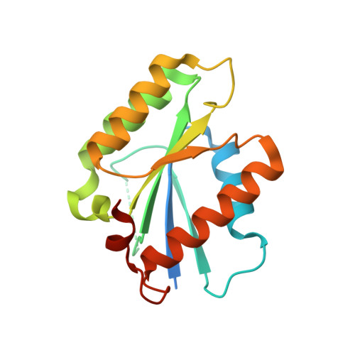
The pseudo GTPase CENP-M drives human kinetochore assembly.
Basilico, F., Maffini, S., Weir, J.R., Prumbaum, D., Rojas, A.M., Zimniak, T., De Antoni, A., Jeganathan, S., Voss, B., van Gerwen, S., Krenn, V., Massimiliano, L., Valencia, A., Vetter, I.R., Herzog, F., Raunser, S., Pasqualato, S., Musacchio, A.(2014) Elife 3: e02978-e02978
- PubMed: 25006165
- DOI: https://doi.org/10.7554/eLife.02978
- Primary Citation of Related Structures:
4P0T - PubMed Abstract:
Kinetochores, multi-subunit complexes that assemble at the interface with centromeres, bind spindle microtubules to ensure faithful delivery of chromosomes during cell division. The configuration and function of the kinetochore-centromere interface is poorly understood. We report that a protein at this interface, CENP-M, is structurally and evolutionarily related to small GTPases but is incapable of GTP-binding and conformational switching. We show that CENP-M is crucially required for the assembly and stability of a tetramer also comprising CENP-I, CENP-H, and CENP-K, the HIKM complex, which we extensively characterize through a combination of structural, biochemical, and cell biological approaches. A point mutant affecting the CENP-M/CENP-I interaction hampers kinetochore assembly and chromosome alignment and prevents kinetochore recruitment of the CENP-T/W complex, questioning a role of CENP-T/W as founder of an independent axis of kinetochore assembly. Our studies identify a single pathway having CENP-C as founder, and CENP-H/I/K/M and CENP-T/W as CENP-C-dependent followers.DOI: http://dx.doi.org/10.7554/eLife.02978.001.
Organizational Affiliation:
Department of Mechanistic Cell Biology, Max Planck Institute of Molecular Physiology, Dortmund, Germany Department of Experimental Oncology, European Institute of Oncology, Milan, Italy.