4P0N

Crystal structure of PDE10a with a novel Imidazo[4,5-b]pyridine inhibitor


Experimental Data Snapshot

  • Method: X-RAY DIFFRACTION
  • Resolution: 2.08 Å
  • R-Value Free: 0.182 
  • R-Value Work: 0.159 
  • R-Value Observed: 0.160 

wwPDB Validation   3D Report Full Report


Ligand Structure Quality Assessment 


This is version 1.1 of the entry. See complete history


Literature

Discovery of Novel Imidazo[4,5-b]pyridines as Potent and Selective Inhibitors of Phosphodiesterase 10A (PDE10A).

Hu, E.Andrews, K.Chmait, S.Zhao, X.Davis, C.Miller, S.Hill Della Puppa, G.Dovlatyan, M.Chen, H.Lester-Zeiner, D.Able, J.Biorn, C.Ma, J.Shi, J.Treanor, J.Allen, J.R.

(2014) ACS Med Chem Lett 5: 700-705

  • DOI: https://doi.org/10.1021/ml5000993
  • Primary Citation of Related Structures:  
    4P0N, 4P1R

  • PubMed Abstract: 

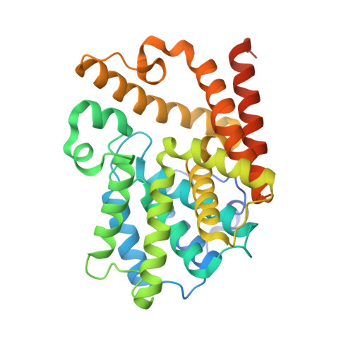
    We report the discovery of novel imidazo[4,5-b]pyridines as potent and selective inhibitors of PDE10A. The investigation began with our recently disclosed ketobenzimidazole 1, which exhibited single digit nanomolar PDE10A activity but poor oral bioavailability. To improve oral bioavailability, we turned to novel scaffold imidazo[4,5-b]pyridine 2, which not only retained nanomolar PDE10A activity but was also devoid of the morpholine metabolic liability. Structure-activity relationship studies were conducted systematically to examine how various regions of the molecule impacted potency. X-ray cocrystal structures of compounds 7 and 24 in human PDE10A helped to elucidate the key bonding interactions. Five of the most potent and structurally diverse imidazo[4,5-b]pyridines (4, 7, 12b, 24a, and 24b) with PDE10A IC50 values ranging from 0.8 to 6.7 nM were advanced into receptor occupancy studies. Four of them (4, 12b, 24a, and 24b) achieved 55-74% RO at 10 mg/kg po.


  • Organizational Affiliation

    Department of Medicinal Chemistry, Department of Molecular Structure, Department of Pharmacokinetics and Drug Metabolism, and Department of Neuroscience, Amgen, Inc. , One Amgen Center Drive, Thousand Oaks, California 93012-1799, United States.


Macromolecules
Find similar proteins by:  (by identity cutoff)  |  3D Structure
Entity ID: 1
MoleculeChains Sequence LengthOrganismDetailsImage
cAMP and cAMP-inhibited cGMP 3',5'-cyclic phosphodiesterase 10A
A, B
338Homo sapiensMutation(s): 0 
Gene Names: PDE10A
EC: 3.1.4.17 (PDB Primary Data), 3.1.4.35 (PDB Primary Data)
UniProt & NIH Common Fund Data Resources
Find proteins for Q9Y233 (Homo sapiens)
Explore Q9Y233 
Go to UniProtKB:  Q9Y233
PHAROS:  Q9Y233
GTEx:  ENSG00000112541 
Entity Groups  
Sequence Clusters30% Identity50% Identity70% Identity90% Identity95% Identity100% Identity
UniProt GroupQ9Y233
Sequence Annotations
Expand
  • Reference Sequence
Small Molecules
Ligands 5 Unique
IDChains Name / Formula / InChI Key2D Diagram3D Interactions
1IS
Query on 1IS

Download Ideal Coordinates CCD File 
C [auth A],
K [auth B]
N-[trans-3-(2-methoxy-3H-imidazo[4,5-b]pyridin-3-yl)cyclobutyl]-1,3-benzothiazol-2-amine
C18 H17 N5 O S
QSZCDYHTHAAFAW-HAQNSBGRSA-N
1IR
Query on 1IR

Download Ideal Coordinates CCD File 
D [auth A],
J [auth B]
N-[cis-3-(2-methoxy-3H-imidazo[4,5-b]pyridin-3-yl)cyclobutyl]-1,3-benzothiazol-2-amine
C18 H17 N5 O S
QSZCDYHTHAAFAW-TXEJJXNPSA-N
SO4
Query on SO4

Download Ideal Coordinates CCD File 
G [auth A]
H [auth A]
N [auth B]
O [auth B]
P [auth B]
G [auth A],
H [auth A],
N [auth B],
O [auth B],
P [auth B],
Q [auth B]
SULFATE ION
O4 S
QAOWNCQODCNURD-UHFFFAOYSA-L
GOL
Query on GOL

Download Ideal Coordinates CCD File 
I [auth A],
R [auth B]
GLYCEROL
C3 H8 O3
PEDCQBHIVMGVHV-UHFFFAOYSA-N
ZN
Query on ZN

Download Ideal Coordinates CCD File 
E [auth A],
F [auth A],
L [auth B],
M [auth B]
ZINC ION
Zn
PTFCDOFLOPIGGS-UHFFFAOYSA-N
Binding Affinity Annotations 
IDSourceBinding Affinity
1IR BindingDB:  4P0N IC50: min: 2.2, max: 3.4 (nM) from 2 assay(s)
Binding MOAD:  4P0N IC50: 3.4 (nM) from 1 assay(s)
1IS Binding MOAD:  4P0N IC50: 0.8 (nM) from 1 assay(s)
BindingDB:  4P0N IC50: 0.8 (nM) from 1 assay(s)
Experimental Data & Validation

Experimental Data

  • Method: X-RAY DIFFRACTION
  • Resolution: 2.08 Å
  • R-Value Free: 0.182 
  • R-Value Work: 0.159 
  • R-Value Observed: 0.160 
  • Space Group: F 2 3
Unit Cell:
Length ( Å )Angle ( ˚ )
a = 253.283α = 90
b = 253.283β = 90
c = 253.283γ = 90
Software Package:
Software NamePurpose
REFMACrefinement

Structure Validation

View Full Validation Report


Ligand Structure Quality Assessment 


Entry History 

Deposition Data

  • Released Date: 2014-10-01 
  • Deposition Author(s): Chmait, S.

Revision History  (Full details and data files)

  • Version 1.0: 2014-10-01
    Type: Initial release
  • Version 1.1: 2023-12-27
    Changes: Data collection, Database references, Derived calculations, Other, Refinement description, Source and taxonomy