4OZU

Crystal Structure of WD40 domain from Toxoplasma gondii coronin


Experimental Data Snapshot

  • Method: X-RAY DIFFRACTION
  • Resolution: 1.65 Å
  • R-Value Free: 0.202 
  • R-Value Work: 0.163 
  • R-Value Observed: 0.165 

wwPDB Validation   3D Report Full Report


This is version 1.5 of the entry. See complete history


Literature

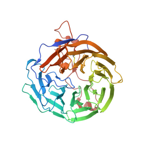
Structure of Toxoplasma gondii coronin, an actin-binding protein that relocalizes to the posterior pole of invasive parasites and contributes to invasion and egress.

Salamun, J.Kallio, J.P.Daher, W.Soldati-Favre, D.Kursula, I.

(2014) FASEB J 28: 4729-4747

  • DOI: https://doi.org/10.1096/fj.14-252569
  • Primary Citation of Related Structures:  
    4OZU

  • PubMed Abstract: 

    Coronins are involved in the regulation of actin dynamics in a multifaceted way, participating in cell migration and vesicular trafficking. Apicomplexan parasites, which exhibit an actin-dependent gliding motility that is essential for traversal through tissues, as well as invasion of and egress from host cells, express only a single coronin, whereas higher eukaryotes possess several isoforms. We set out to characterize the 3-D structure, biochemical function, subcellular localization, and genetic ablation of Toxoplasma gondii coronin (TgCOR), to shed light on its biological role. A combination of X-ray crystallography, small-angle scattering of X-rays, and light scattering revealed the atomic structure of the conserved WD40 domain and the dimeric arrangement of the full-length protein. TgCOR binds to F-actin and increases the rate and extent of actin polymerization. In vivo, TgCOR relocalizes transiently to the posterior pole of motile and invading parasites, independent of actin dynamics, but concomitant to microneme secretory organelle discharge. TgCOR contributes to, but is not essential for, invasion and egress. Taken together, our data point toward a role for TgCOR in stabilizing newly formed, short filaments and F-actin cross-linking, as well as functions linked to endocytosis and recycling of membranes.


  • Organizational Affiliation

    Department of Microbiology and Molecular Medicine, Faculty of Medicine, University of Geneva, Geneva, Switzerland;


Macromolecules
Find similar proteins by:  (by identity cutoff)  |  3D Structure
Entity ID: 1
MoleculeChains Sequence LengthOrganismDetailsImage
Coronin394Toxoplasma gondiiMutation(s): 0 
Gene Names: CRN1
UniProt
Find proteins for Q5Y1E7 (Toxoplasma gondii)
Explore Q5Y1E7 
Go to UniProtKB:  Q5Y1E7
Entity Groups  
Sequence Clusters30% Identity50% Identity70% Identity90% Identity95% Identity100% Identity
UniProt GroupQ5Y1E7
Sequence Annotations
Expand
  • Reference Sequence
Small Molecules
Ligands 2 Unique
IDChains Name / Formula / InChI Key2D Diagram3D Interactions
TRS
Query on TRS

Download Ideal Coordinates CCD File 
B [auth A]2-AMINO-2-HYDROXYMETHYL-PROPANE-1,3-DIOL
C4 H12 N O3
LENZDBCJOHFCAS-UHFFFAOYSA-O
ACT
Query on ACT

Download Ideal Coordinates CCD File 
C [auth A]ACETATE ION
C2 H3 O2
QTBSBXVTEAMEQO-UHFFFAOYSA-M
Experimental Data & Validation

Experimental Data

  • Method: X-RAY DIFFRACTION
  • Resolution: 1.65 Å
  • R-Value Free: 0.202 
  • R-Value Work: 0.163 
  • R-Value Observed: 0.165 
  • Space Group: C 2 2 21
Unit Cell:
Length ( Å )Angle ( ˚ )
a = 55.13α = 90
b = 82.51β = 90
c = 156.98γ = 90
Software Package:
Software NamePurpose
PHENIXrefinement

Structure Validation

View Full Validation Report


Entry History 

Deposition Data

Revision History  (Full details and data files)

  • Version 1.0: 2014-08-20
    Type: Initial release
  • Version 1.1: 2014-08-27
    Changes: Database references
  • Version 1.2: 2014-11-12
    Changes: Database references
  • Version 1.3: 2015-02-04
    Changes: Derived calculations
  • Version 1.4: 2018-03-07
    Changes: Advisory, Data collection, Database references, Derived calculations, Other, Source and taxonomy
  • Version 1.5: 2023-09-27
    Changes: Data collection, Database references, Refinement description