4OYU

Crystal structure of the N-terminal domains of muskelin


Experimental Data Snapshot

  • Method: X-RAY DIFFRACTION
  • Resolution: 1.80 Å
  • R-Value Free: 0.193 
  • R-Value Work: 0.156 
  • R-Value Observed: 0.158 

wwPDB Validation   3D Report Full Report


This is version 1.3 of the entry. See complete history


Literature

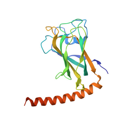
The LisH Motif of Muskelin Is Crucial for Oligomerization and Governs Intracellular Localization.

Delto, C.F.Heisler, F.F.Kuper, J.Sander, B.Kneussel, M.Schindelin, H.

(2015) Structure 23: 364-373

  • DOI: https://doi.org/10.1016/j.str.2014.11.016
  • Primary Citation of Related Structures:  
    4OYU

  • PubMed Abstract: 

    Neurons regulate the number of surface receptors by balancing the transport to and from the plasma membrane to adjust their signaling properties. The protein muskelin was recently identified as a key factor guiding the transport of α1 subunit-containing GABAA receptors. Here we present the crystal structure of muskelin, comprising its N-terminal discoidin domain and Lis1-homology (LisH) motif. The molecule crystallized as a dimer with the LisH motif exclusively mediating oligomerization. Our subsequent biochemical analyses confirmed that the LisH motif acts as a dimerization element in muskelin. Together with an intermolecular head-to-tail interaction, the LisH-dependent dimerization is required to assemble a muskelin tetramer. Intriguingly, our cellular studies revealed that the loss of this dimerization results in a complete redistribution of muskelin from the cytoplasm to the nucleus and impairs muskelin's function in GABAA receptor transport. These studies demonstrate that the LisH-dependent dimerization is a crucial factor for muskelin function.


  • Organizational Affiliation

    Rudolf Virchow Center for Experimental Biomedicine, University of Würzburg, D-97080 Würzburg, Germany.


Macromolecules
Find similar proteins by:  (by identity cutoff)  |  3D Structure
Entity ID: 1
MoleculeChains Sequence LengthOrganismDetailsImage
Muskelin
A, B
196Rattus norvegicusMutation(s): 0 
Gene Names: Mkln1Msk
UniProt
Find proteins for Q99PV3 (Rattus norvegicus)
Explore Q99PV3 
Go to UniProtKB:  Q99PV3
Entity Groups  
Sequence Clusters30% Identity50% Identity70% Identity90% Identity95% Identity100% Identity
UniProt GroupQ99PV3
Sequence Annotations
Expand
  • Reference Sequence
Small Molecules
Ligands 2 Unique
IDChains Name / Formula / InChI Key2D Diagram3D Interactions
GOL
Query on GOL

Download Ideal Coordinates CCD File 
M [auth A]GLYCEROL
C3 H8 O3
PEDCQBHIVMGVHV-UHFFFAOYSA-N
EDO
Query on EDO

Download Ideal Coordinates CCD File 
C [auth A]
D [auth A]
E [auth A]
F [auth A]
G [auth A]
C [auth A],
D [auth A],
E [auth A],
F [auth A],
G [auth A],
H [auth A],
I [auth A],
J [auth A],
K [auth A],
L [auth A],
N [auth A],
O [auth A],
P [auth A],
Q [auth B],
R [auth B],
S [auth B],
T [auth B],
U [auth B],
V [auth B],
W [auth B],
X [auth B],
Y [auth B],
Z [auth B]
1,2-ETHANEDIOL
C2 H6 O2
LYCAIKOWRPUZTN-UHFFFAOYSA-N
Experimental Data & Validation

Experimental Data

  • Method: X-RAY DIFFRACTION
  • Resolution: 1.80 Å
  • R-Value Free: 0.193 
  • R-Value Work: 0.156 
  • R-Value Observed: 0.158 
  • Space Group: P 21 21 21
Unit Cell:
Length ( Å )Angle ( ˚ )
a = 63.06α = 90
b = 65.3β = 90
c = 101.39γ = 90
Software Package:
Software NamePurpose
PHENIXrefinement
PHASERphasing
SCALAdata scaling
MOSFLMdata reduction
PDB_EXTRACTdata extraction

Structure Validation

View Full Validation Report


Entry History 

Deposition Data

Revision History  (Full details and data files)

  • Version 1.0: 2015-02-11
    Type: Initial release
  • Version 1.1: 2015-02-18
    Changes: Database references, Derived calculations
  • Version 1.2: 2015-03-25
    Changes: Derived calculations
  • Version 1.3: 2023-12-27
    Changes: Data collection, Database references, Derived calculations, Source and taxonomy