4OYM

Human solAC Complexed with (4-Amino-furazan-3-yl)-(3-methoxy-phenyl)-methanone


Experimental Data Snapshot

  • Method: X-RAY DIFFRACTION
  • Resolution: 1.70 Å
  • R-Value Free: 0.218 
  • R-Value Work: 0.174 
  • R-Value Observed: 0.176 

wwPDB Validation   3D Report Full Report


Ligand Structure Quality Assessment 


This is version 1.2 of the entry. See complete history


Literature

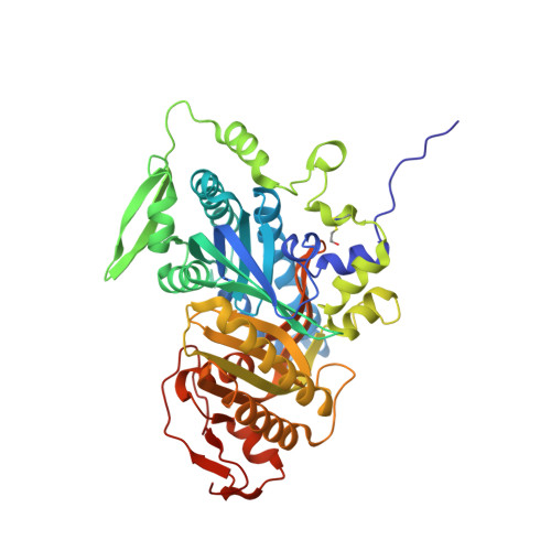
Crystal structure of human soluble adenylate cyclase reveals a distinct, highly flexible allosteric bicarbonate binding pocket.

Saalau-Bethell, S.M.Berdini, V.Cleasby, A.Congreve, M.Coyle, J.E.Lock, V.Murray, C.W.O'Brien, M.A.Rich, S.J.Sambrook, T.Vinkovic, M.Yon, J.R.Jhoti, H.

(2014) ChemMedChem 9: 823-832

  • DOI: https://doi.org/10.1002/cmdc.201300480
  • Primary Citation of Related Structures:  
    4OYA, 4OYB, 4OYI, 4OYM, 4OYO, 4OYP, 4OYW, 4OYX, 4OYZ, 4OZ2, 4OZ3

  • PubMed Abstract: 

    Soluble adenylate cyclases catalyse the synthesis of the second messenger cAMP through the cyclisation of ATP and are the only known enzymes to be directly activated by bicarbonate. Here, we report the first crystal structure of the human enzyme that reveals a pseudosymmetrical arrangement of two catalytic domains to produce a single competent active site and a novel discrete bicarbonate binding pocket. Crystal structures of the apo protein, the protein in complex with α,β-methylene adenosine 5'-triphosphate (AMPCPP) and calcium, with the allosteric activator bicarbonate, and also with a number of inhibitors identified using fragment screening, all show a flexible active site that undergoes significant conformational changes on binding of ligands. The resulting nanomolar-potent inhibitors that were developed bind at both the substrate binding pocket and the allosteric site, and can be used as chemical probes to further elucidate the function of this protein.


  • Organizational Affiliation

    Astex Pharmaceuticals, Cambridge Science Park, Cambridge, CB4 0QA (UK).


Macromolecules
Find similar proteins by:  (by identity cutoff)  |  3D Structure
Entity ID: 1
MoleculeChains Sequence LengthOrganismDetailsImage
Adenylate cyclase type 10470Homo sapiensMutation(s): 1 
Gene Names: ADCY10SAC
EC: 4.6.1.1
UniProt & NIH Common Fund Data Resources
Find proteins for Q96PN6 (Homo sapiens)
Explore Q96PN6 
Go to UniProtKB:  Q96PN6
PHAROS:  Q96PN6
GTEx:  ENSG00000143199 
Entity Groups  
Sequence Clusters30% Identity50% Identity70% Identity90% Identity95% Identity100% Identity
UniProt GroupQ96PN6
Sequence Annotations
Expand
  • Reference Sequence
Small Molecules
Modified Residues  1 Unique
IDChains TypeFormula2D DiagramParent
CME
Query on CME
A
L-PEPTIDE LINKINGC5 H11 N O3 S2CYS
Binding Affinity Annotations 
IDSourceBinding Affinity
1ZC Binding MOAD:  4OYM IC50: 1.10e+4 (nM) from 1 assay(s)
Experimental Data & Validation

Experimental Data

  • Method: X-RAY DIFFRACTION
  • Resolution: 1.70 Å
  • R-Value Free: 0.218 
  • R-Value Work: 0.174 
  • R-Value Observed: 0.176 
  • Space Group: P 63
Unit Cell:
Length ( Å )Angle ( ˚ )
a = 99.343α = 90
b = 99.343β = 90
c = 97.964γ = 120
Software Package:
Software NamePurpose
BUSTERrefinement

Structure Validation

View Full Validation Report


Ligand Structure Quality Assessment 


Entry History 

Deposition Data

  • Released Date: 2014-04-02 
  • Deposition Author(s): Vinkovic, M.

Revision History  (Full details and data files)

  • Version 1.0: 2014-04-02
    Type: Initial release
  • Version 1.1: 2014-10-01
    Changes: Database references
  • Version 1.2: 2023-12-27
    Changes: Data collection, Database references, Derived calculations, Other, Refinement description, Source and taxonomy