4OWM

Anthranilate phosphoribosyl transferase from Mycobacterium tuberculosis in complex with 3-fluoroanthranilate, PRPP and Magnesium


Experimental Data Snapshot

  • Method: X-RAY DIFFRACTION
  • Resolution: 1.99 Å
  • R-Value Free: 0.213 
  • R-Value Work: 0.170 
  • R-Value Observed: 0.172 

wwPDB Validation   3D Report Full Report


Ligand Structure Quality Assessment 


This is version 1.6 of the entry. See complete history


Literature

Alternative substrates reveal catalytic cycle and key binding events in the reaction catalysed by anthranilate phosphoribosyltransferase from Mycobacterium tuberculosis.

Cookson, T.V.Castell, A.Bulloch, E.M.Evans, G.L.Short, F.L.Baker, E.N.Lott, J.S.Parker, E.J.

(2014) Biochem J 461: 87-98

  • DOI: https://doi.org/10.1042/BJ20140209
  • Primary Citation of Related Structures:  
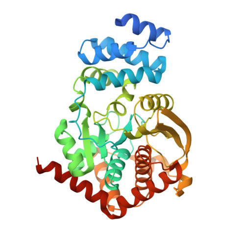
    4N5V, 4N8Q, 4N93, 4OWM, 4OWN, 4OWO, 4OWQ, 4OWS, 4OWU, 4OWV

  • PubMed Abstract: 

    AnPRT (anthranilate phosphoribosyltransferase), required for the biosynthesis of tryptophan, is essential for the virulence of Mycobacterium tuberculosis (Mtb). AnPRT catalyses the Mg2+-dependent transfer of a phosphoribosyl group from PRPP (5'-phosphoribosyl-1'-pyrophosphate) to anthranilate to form PRA (5'-phosphoribosyl anthranilate). Mtb-AnPRT was shown to catalyse a sequential reaction and significant substrate inhibition by anthranilate was observed. Antimycobacterial fluoroanthranilates and methyl-substituted analogues were shown to act as alternative substrates for Mtb-AnPRT, producing the corresponding substituted PRA products. Structures of the enzyme complexed with anthranilate analogues reveal two distinct binding sites for anthranilate. One site is located over 8 Å (1 Å=0.1 nm) from PRPP at the entrance to a tunnel leading to the active site, whereas in the second, inner, site anthranilate is adjacent to PRPP, in a catalytically relevant position. Soaking the analogues for variable periods of time provides evidence for anthranilate located at transient positions during transfer from the outer site to the inner catalytic site. PRPP and Mg2+ binding have been shown to be associated with the rearrangement of two flexible loops, which is required to complete the inner anthranilate-binding site. It is proposed that anthranilate first binds to the outer site, providing an unusual mechanism for substrate capture and efficient transfer to the catalytic site following the binding of PRPP.


  • Organizational Affiliation

    *Maurice Wilkins Centre for Molecular Biodiscovery, Biomolecular Interaction Centre and Department of Chemistry, University of Canterbury, 20 Kirkwood Avenue, Christchurch 8140, New Zealand.


Macromolecules
Find similar proteins by:  (by identity cutoff)  |  3D Structure
Entity ID: 1
MoleculeChains Sequence LengthOrganismDetailsImage
Anthranilate phosphoribosyltransferase
A, B
378Mycobacterium tuberculosisMutation(s): 0 
Gene Names: MT2248MTCY190.03cRv2192ctrpD
EC: 2.4.2.18
UniProt
Find proteins for P9WFX5 (Mycobacterium tuberculosis (strain ATCC 25618 / H37Rv))
Explore P9WFX5 
Go to UniProtKB:  P9WFX5
Entity Groups  
Sequence Clusters30% Identity50% Identity70% Identity90% Identity95% Identity100% Identity
UniProt GroupP9WFX5
Sequence Annotations
Expand
  • Reference Sequence
Small Molecules
Ligands 5 Unique
IDChains Name / Formula / InChI Key2D Diagram3D Interactions
PRP
Query on PRP

Download Ideal Coordinates CCD File 
E [auth A],
J [auth B]
1-O-pyrophosphono-5-O-phosphono-alpha-D-ribofuranose
C5 H13 O14 P3
PQGCEDQWHSBAJP-TXICZTDVSA-N
3F0
Query on 3F0

Download Ideal Coordinates CCD File 
F [auth A],
K [auth B]
2-azanyl-3-fluoranyl-benzoic acid
C7 H6 F N O2
KUHAYJJXXGBYBW-UHFFFAOYSA-N
GOL
Query on GOL

Download Ideal Coordinates CCD File 
G [auth A]GLYCEROL
C3 H8 O3
PEDCQBHIVMGVHV-UHFFFAOYSA-N
IMD
Query on IMD

Download Ideal Coordinates CCD File 
L [auth B]IMIDAZOLE
C3 H5 N2
RAXXELZNTBOGNW-UHFFFAOYSA-O
MG
Query on MG

Download Ideal Coordinates CCD File 
C [auth A],
D [auth A],
H [auth B],
I [auth B]
MAGNESIUM ION
Mg
JLVVSXFLKOJNIY-UHFFFAOYSA-N
Experimental Data & Validation

Experimental Data

  • Method: X-RAY DIFFRACTION
  • Resolution: 1.99 Å
  • R-Value Free: 0.213 
  • R-Value Work: 0.170 
  • R-Value Observed: 0.172 
  • Space Group: P 21 21 21
Unit Cell:
Length ( Å )Angle ( ˚ )
a = 79.274α = 90
b = 91.99β = 90
c = 120.892γ = 90
Software Package:
Software NamePurpose
SCALAdata scaling
REFMACrefinement
PDB_EXTRACTdata extraction

Structure Validation

View Full Validation Report


Ligand Structure Quality Assessment 


Entry History 

Deposition Data

Revision History  (Full details and data files)

  • Version 1.0: 2014-04-23
    Type: Initial release
  • Version 1.1: 2014-10-01
    Changes: Database references
  • Version 1.2: 2014-12-17
    Changes: Data collection
  • Version 1.3: 2017-09-27
    Changes: Data collection, Database references, Derived calculations, Other, Refinement description, Source and taxonomy
  • Version 1.4: 2018-03-07
    Changes: Data collection
  • Version 1.5: 2020-07-29
    Type: Remediation
    Reason: Carbohydrate remediation
    Changes: Data collection, Derived calculations, Refinement description, Structure summary
  • Version 1.6: 2023-12-27
    Changes: Data collection, Database references, Structure summary