4OUC

Structure of human haspin in complex with histone H3 substrate


Experimental Data Snapshot

  • Method: X-RAY DIFFRACTION
  • Resolution: 1.90 Å
  • R-Value Free: 0.211 
  • R-Value Work: 0.164 
  • R-Value Observed: 0.166 

wwPDB Validation   3D Report Full Report


Ligand Structure Quality Assessment 


This is version 1.2 of the entry. See complete history


Literature

Modulation of the chromatin phosphoproteome by the haspin protein kinase.

Maiolica, A.de Medina-Redondo, M.Schoof, E.M.Chaikuad, A.Villa, F.Gatti, M.Jeganathan, S.Lou, H.J.Novy, K.Hauri, S.Toprak, U.H.Herzog, F.Meraldi, P.Penengo, L.Turk, B.E.Knapp, S.Linding, R.Aebersold, R.

(2014) Mol Cell Proteomics 13: 1724-1740

  • DOI: https://doi.org/10.1074/mcp.M113.034819
  • Primary Citation of Related Structures:  
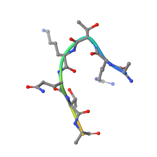
    4OUC

  • PubMed Abstract: 

    Recent discoveries have highlighted the importance of Haspin kinase activity for the correct positioning of the kinase Aurora B at the centromere. Haspin phosphorylates Thr(3) of the histone H3 (H3), which provides a signal for Aurora B to localize to the centromere of mitotic chromosomes. To date, histone H3 is the only confirmed Haspin substrate. We used a combination of biochemical, pharmacological, and mass spectrometric approaches to study the consequences of Haspin inhibition in mitotic cells. We quantified 3964 phosphorylation sites on chromatin-associated proteins and identified a Haspin protein-protein interaction network. We determined the Haspin consensus motif and the co-crystal structure of the kinase with the histone H3 tail. The structure revealed a unique bent substrate binding mode positioning the histone H3 residues Arg(2) and Lys(4) adjacent to the Haspin phosphorylated threonine into acidic binding pockets. This unique conformation of the kinase-substrate complex explains the reported modulation of Haspin activity by methylation of Lys(4) of the histone H3. In addition, the identification of the structural basis of substrate recognition and the amino acid sequence preferences of Haspin aided the identification of novel candidate Haspin substrates. In particular, we validated the phosphorylation of Ser(137) of the histone variant macroH2A as a target of Haspin kinase activity. MacroH2A Ser(137) resides in a basic stretch of about 40 amino acids that is required to stabilize extranucleosomal DNA, suggesting that phosphorylation of Ser(137) might regulate the interactions of macroH2A and DNA. Overall, our data suggest that Haspin activity affects the phosphorylation state of proteins involved in gene expression regulation and splicing.


  • Organizational Affiliation

    From the ‡Department of Biology, Institute of Molecular Systems Biology, ETH Zurich, Zurich, Switzerland;


Macromolecules
Find similar proteins by:  (by identity cutoff)  |  3D Structure
Entity ID: 1
MoleculeChains Sequence LengthOrganismDetailsImage
Serine/threonine-protein kinase haspin357Homo sapiensMutation(s): 0 
Gene Names: GSG2
EC: 2.7.11.1
UniProt & NIH Common Fund Data Resources
Find proteins for Q8TF76 (Homo sapiens)
Explore Q8TF76 
Go to UniProtKB:  Q8TF76
PHAROS:  Q8TF76
GTEx:  ENSG00000177602 
Entity Groups  
Sequence Clusters30% Identity50% Identity70% Identity90% Identity95% Identity100% Identity
UniProt GroupQ8TF76
Sequence Annotations
Expand
  • Reference Sequence

Find similar proteins by:  Sequence   |   3D Structure  

Entity ID: 2
MoleculeChains Sequence LengthOrganismDetailsImage
Histone H3.212Homo sapiensMutation(s): 0 
UniProt & NIH Common Fund Data Resources
Find proteins for Q71DI3 (Homo sapiens)
Explore Q71DI3 
Go to UniProtKB:  Q71DI3
PHAROS:  Q71DI3
Entity Groups  
Sequence Clusters30% Identity50% Identity70% Identity90% Identity95% Identity100% Identity
UniProt GroupQ71DI3
Sequence Annotations
Expand
  • Reference Sequence
Small Molecules
Ligands 4 Unique
IDChains Name / Formula / InChI Key2D Diagram3D Interactions
5ID
Query on 5ID

Download Ideal Coordinates CCD File 
J [auth A](2R,3R,4S,5R)-2-(4-AMINO-5-IODO-7H-PYRROLO[2,3-D]PYRIMIDIN-7-YL)-5-(HYDROXYMETHYL)TETRAHYDROFURAN-3,4-DIOL
C11 H13 I N4 O4
WHSIXKUPQCKWBY-IOSLPCCCSA-N
IOD
Query on IOD

Download Ideal Coordinates CCD File 
K [auth A]IODIDE ION
I
XMBWDFGMSWQBCA-UHFFFAOYSA-M
EDO
Query on EDO

Download Ideal Coordinates CCD File 
D [auth A]
E [auth A]
F [auth A]
G [auth A]
H [auth A]
D [auth A],
E [auth A],
F [auth A],
G [auth A],
H [auth A],
I [auth A]
1,2-ETHANEDIOL
C2 H6 O2
LYCAIKOWRPUZTN-UHFFFAOYSA-N
NA
Query on NA

Download Ideal Coordinates CCD File 
C [auth A]SODIUM ION
Na
FKNQFGJONOIPTF-UHFFFAOYSA-N
Experimental Data & Validation

Experimental Data

  • Method: X-RAY DIFFRACTION
  • Resolution: 1.90 Å
  • R-Value Free: 0.211 
  • R-Value Work: 0.164 
  • R-Value Observed: 0.166 
  • Space Group: P 21 21 21
Unit Cell:
Length ( Å )Angle ( ˚ )
a = 50.54α = 90
b = 79.13β = 90
c = 100.76γ = 90
Software Package:
Software NamePurpose
CrystalCleardata collection
PHASERphasing
REFMACrefinement
MOSFLMdata reduction
SCALAdata scaling

Structure Validation

View Full Validation Report


Ligand Structure Quality Assessment 


Entry History 

Deposition Data

Revision History  (Full details and data files)

  • Version 1.0: 2014-04-16
    Type: Initial release
  • Version 1.1: 2014-08-13
    Changes: Database references
  • Version 1.2: 2023-09-20
    Changes: Data collection, Database references, Derived calculations, Refinement description