4OR7

Klebsiella pneumoniae dihydrofolate reductase complexed with NADPH and 6-ethyl-5-{3-[3-(pyrimidin-5-yl)phenyl]prop-1-yn-1-yl}pyrimidine-2,4-diamine


Experimental Data Snapshot

  • Method: X-RAY DIFFRACTION
  • Resolution: 1.76 Å
  • R-Value Free: 0.250 
  • R-Value Work: 0.210 
  • R-Value Observed: 0.213 

wwPDB Validation   3D Report Full Report


Ligand Structure Quality Assessment 


This is version 1.2 of the entry. See complete history


Literature

Crystal Structures of Klebsiella pneumoniae Dihydrofolate Reductase Bound to Propargyl-Linked Antifolates Reveal Features for Potency and Selectivity.

Lamb, K.M.Lombardo, M.N.Alverson, J.Priestley, N.D.Wright, D.L.Anderson, A.C.

(2014) Antimicrob Agents Chemother 58: 7484-7491

  • DOI: https://doi.org/10.1128/AAC.03555-14
  • Primary Citation of Related Structures:  
    4OR7, 4OSG

  • PubMed Abstract: 

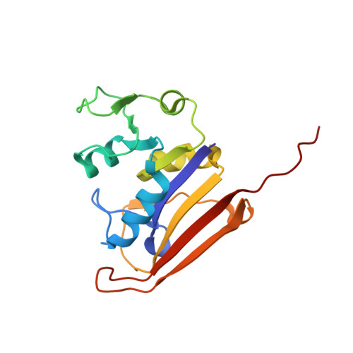
    Resistance to the antibacterial antifolate trimethoprim (TMP) is increasing in members of the family Enterobacteriaceae, driving the design of next-generation antifolates effective against these Gram-negative pathogens. The propargyl-linked antifolates are potent inhibitors of dihydrofolate reductases (DHFR) from several TMP-sensitive and -resistant species, including Klebsiella pneumoniae. Recently, we have determined that these antifolates inhibit the growth of strains of K. pneumoniae, some with MIC values of 1 μg/ml. In order to further the design of potent and selective antifolates against members of the Enterobacteriaceae, we determined the first crystal structures of K. pneumoniae DHFR bound to two of the propargyl-linked antifolates. These structures highlight that interactions with Leu 28, Ile 50, Ile 94, and Leu 54 are necessary for potency; comparison with structures of human DHFR bound to the same inhibitors reveal differences in residues (N64E, P61G, F31L, and V115I) and loop conformations (residues 49 to 53) that may be exploited for selectivity.


  • Organizational Affiliation

    Department of Pharmaceutical Sciences, University of Connecticut, Storrs, Connecticut, USA.


Macromolecules
Find similar proteins by:  (by identity cutoff)  |  3D Structure
Entity ID: 1
MoleculeChains Sequence LengthOrganismDetailsImage
Dihydrofolate reductase165Klebsiella pneumoniae CG43Mutation(s): 0 
Gene Names: folAD364_00170
EC: 1.5.1.3
Entity Groups  
Sequence Clusters30% Identity50% Identity70% Identity90% Identity95% Identity100% Identity
Sequence Annotations
Expand
  • Reference Sequence
Small Molecules
Ligands 2 Unique
IDChains Name / Formula / InChI Key2D Diagram3D Interactions
NAP
Query on NAP

Download Ideal Coordinates CCD File 
B [auth A]NADP NICOTINAMIDE-ADENINE-DINUCLEOTIDE PHOSPHATE
C21 H28 N7 O17 P3
XJLXINKUBYWONI-NNYOXOHSSA-N
25U
Query on 25U

Download Ideal Coordinates CCD File 
C [auth A]6-ethyl-5-{3-[3-(pyrimidin-5-yl)phenyl]prop-1-yn-1-yl}pyrimidine-2,4-diamine
C19 H18 N6
UEZKDWYIIOOENX-UHFFFAOYSA-N
Binding Affinity Annotations 
IDSourceBinding Affinity
25U Binding MOAD:  4OR7 IC50: 23 (nM) from 1 assay(s)
Experimental Data & Validation

Experimental Data

  • Method: X-RAY DIFFRACTION
  • Resolution: 1.76 Å
  • R-Value Free: 0.250 
  • R-Value Work: 0.210 
  • R-Value Observed: 0.213 
  • Space Group: P 32 2 1
Unit Cell:
Length ( Å )Angle ( ˚ )
a = 61.161α = 90
b = 61.161β = 90
c = 105.426γ = 120
Software Package:
Software NamePurpose
HKL-2000data collection
PHASERphasing
PHENIXrefinement
HKL-2000data reduction
d*TREKdata scaling

Structure Validation

View Full Validation Report


Ligand Structure Quality Assessment 


Entry History 

Deposition Data

Revision History  (Full details and data files)

  • Version 1.0: 2014-12-03
    Type: Initial release
  • Version 1.1: 2014-12-17
    Changes: Data collection
  • Version 1.2: 2024-02-28
    Changes: Data collection, Database references, Derived calculations