4OOE

M. tuberculosis 1-deoxy-d-xylulose-5-phosphate reductoisomerase W203Y mutant bound to fosmidomycin and NADPH


Experimental Data Snapshot

  • Method: X-RAY DIFFRACTION
  • Resolution: 1.83 Å
  • R-Value Free: 0.211 
  • R-Value Work: 0.170 
  • R-Value Observed: 0.170 

wwPDB Validation   3D Report Full Report


Ligand Structure Quality Assessment 


This is version 1.1 of the entry. See complete history


Literature

Alteration of the Flexible Loop in 1-Deoxy-d-xylulose-5-phosphate Reductoisomerase Boosts Enthalpy-Driven Inhibition by Fosmidomycin.

Kholodar, S.A.Tombline, G.Liu, J.Tan, Z.Allen, C.L.Gulick, A.M.Murkin, A.S.

(2014) Biochemistry 53: 3423-3431

  • DOI: https://doi.org/10.1021/bi5004074
  • Primary Citation of Related Structures:  
    4OOE, 4OOF

  • PubMed Abstract: 

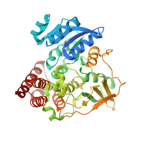
    1-Deoxy-d-xylulose-5-phosphate reductoisomerase (DXR), which catalyzes the first committed step in the 2-C-methyl-d-erythritol 4-phosphate pathway of isoprenoid biosynthesis used by Mycobacterium tuberculosis and other infectious microorganisms, is absent in humans and therefore an attractive drug target. Fosmidomycin is a nanomolar inhibitor of DXR, but despite great efforts, few analogues with comparable potency have been developed. DXR contains a strictly conserved residue, Trp203, within a flexible loop that closes over and interacts with the bound inhibitor. We report that while mutation to Ala or Gly abolishes activity, mutation to Phe and Tyr only modestly impacts kcat and Km. Moreover, pre-steady-state kinetics and primary deuterium kinetic isotope effects indicate that while turnover is largely limited by product release for the wild-type enzyme, chemistry is significantly more rate-limiting for W203F and W203Y. Surprisingly, these mutants are more sensitive to inhibition by fosmidomycin, resulting in Km/Ki ratios up to 19-fold higher than that of wild-type DXR. In agreement, isothermal titration calorimetry revealed that fosmidomycin binds up to 11-fold more tightly to these mutants. Most strikingly, mutation strongly tips the entropy-enthalpy balance of total binding energy from 50% to 75% and 91% enthalpy in W203F and W203Y, respectively. X-ray crystal structures suggest that these enthalpy differences may be linked to differences in hydrogen bond interactions involving a water network connecting fosmidomycin's phosphonate group to the protein. These results confirm the importance of the flexible loop, in particular Trp203, in ligand binding and suggest that improved inhibitor affinity may be obtained against the wild-type protein by introducing interactions with this loop and/or the surrounding structured water network.


  • Organizational Affiliation

    Department of Chemistry, University at Buffalo , Buffalo, New York 14260-3000, United States.


Macromolecules
Find similar proteins by:  (by identity cutoff)  |  3D Structure
Entity ID: 1
MoleculeChains Sequence LengthOrganismDetailsImage
1-deoxy-D-xylulose 5-phosphate reductoisomerase
A, B, C, D
404Mycobacterium tuberculosis H37RvMutation(s): 1 
Gene Names: dxrRVBD_2870c
EC: 1.1.1.267
UniProt
Find proteins for P9WNS1 (Mycobacterium tuberculosis (strain ATCC 25618 / H37Rv))
Explore P9WNS1 
Go to UniProtKB:  P9WNS1
Entity Groups  
Sequence Clusters30% Identity50% Identity70% Identity90% Identity95% Identity100% Identity
UniProt GroupP9WNS1
Sequence Annotations
Expand
  • Reference Sequence
Small Molecules
Ligands 3 Unique
IDChains Name / Formula / InChI Key2D Diagram3D Interactions
NDP
Query on NDP

Download Ideal Coordinates CCD File 
G [auth A],
J [auth B],
M [auth C],
P [auth D]
NADPH DIHYDRO-NICOTINAMIDE-ADENINE-DINUCLEOTIDE PHOSPHATE
C21 H30 N7 O17 P3
ACFIXJIJDZMPPO-NNYOXOHSSA-N
FOM
Query on FOM

Download Ideal Coordinates CCD File 
E [auth A],
H [auth B],
K [auth C],
N [auth D]
3-[FORMYL(HYDROXY)AMINO]PROPYLPHOSPHONIC ACID
C4 H10 N O5 P
GJXWDTUCERCKIX-UHFFFAOYSA-N
MN
Query on MN

Download Ideal Coordinates CCD File 
F [auth A],
I [auth B],
L [auth C],
O [auth D]
MANGANESE (II) ION
Mn
WAEMQWOKJMHJLA-UHFFFAOYSA-N
Binding Affinity Annotations 
IDSourceBinding Affinity
FOM BindingDB:  4OOE Ki: 140 (nM) from 1 assay(s)
Kd: 40 (nM) from 1 assay(s)
IC50: min: 80, max: 440 (nM) from 3 assay(s)
Binding MOAD:  4OOE Ki: 14 (nM) from 1 assay(s)
Experimental Data & Validation

Experimental Data

  • Method: X-RAY DIFFRACTION
  • Resolution: 1.83 Å
  • R-Value Free: 0.211 
  • R-Value Work: 0.170 
  • R-Value Observed: 0.170 
  • Space Group: P 21 21 21
Unit Cell:
Length ( Å )Angle ( ˚ )
a = 112.27α = 90
b = 114.24β = 90
c = 133.187γ = 90
Software Package:
Software NamePurpose
Blu-Icedata collection
PHASERphasing
PHENIXrefinement
XDSdata reduction
SCALAdata scaling

Structure Validation

View Full Validation Report


Ligand Structure Quality Assessment 


Entry History 

Deposition Data

Revision History  (Full details and data files)

  • Version 1.0: 2014-06-18
    Type: Initial release
  • Version 1.1: 2023-09-20
    Changes: Data collection, Database references, Derived calculations, Refinement description