4OL0

Crystal structure of transportin-SR2, a karyopherin involved in human disease, in complex with Ran


Experimental Data Snapshot

  • Method: X-RAY DIFFRACTION
  • Resolution: 2.90 Å
  • R-Value Free: 0.276 
  • R-Value Work: 0.222 
  • R-Value Observed: 0.224 

wwPDB Validation   3D Report Full Report


Ligand Structure Quality Assessment 


This is version 1.2 of the entry. See complete history


Literature

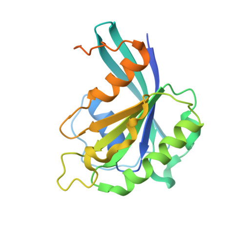
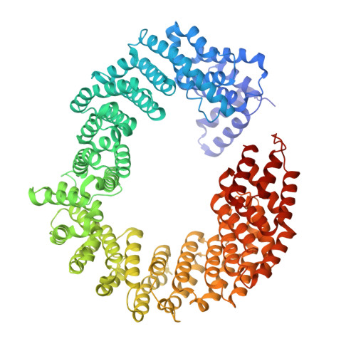
Structure of transportin SR2, a karyopherin involved in human disease, in complex with Ran.

Tsirkone, V.G.Beutels, K.G.Demeulemeester, J.Debyser, Z.Christ, F.Strelkov, S.V.

(2014) Acta Crystallogr Sect F Struct Biol Cryst Commun 70: 723-729

  • DOI: https://doi.org/10.1107/S2053230X14009492
  • Primary Citation of Related Structures:  
    4OL0

  • PubMed Abstract: 

    Transportin SR2 (TRN-SR2) is a β-type karyopherin responsible for the nuclear import of specific cargoes, including serine/arginine-rich splicing factors. The protein has been implicated in a variety of human diseases, including HIV infection, primary biliary cirrhosis and limb-girdle muscular dystrophy 1F. Towards understanding its molecular mechanism, a 2.9 Å resolution crystal structure of human TRN-SR2 complexed with the small GTPase Ran has been determined. TRN-SR2 is composed of 20 α-helical HEAT repeats forming a solenoid-like fold. The first nine repeats form a `cradle' for the binding of RanGTP, revealing similarities but also differences with respect to the related importin 13 complex.


  • Organizational Affiliation

    Laboratory for Biocrystallography, Department of Pharmaceutical and Pharmacological Sciences, KU Leuven, Herestraat 49 bus 822, 3000 Leuven, Belgium.


Macromolecules
Find similar proteins by:  (by identity cutoff)  |  3D Structure
Entity ID: 1
MoleculeChains Sequence LengthOrganismDetailsImage
GTP-binding nuclear protein Ran216Homo sapiensMutation(s): 0 
Gene Names: RANARA24OK/SW-cl.81
UniProt & NIH Common Fund Data Resources
Find proteins for P62826 (Homo sapiens)
Explore P62826 
Go to UniProtKB:  P62826
PHAROS:  P62826
GTEx:  ENSG00000132341 
Entity Groups  
Sequence Clusters30% Identity50% Identity70% Identity90% Identity95% Identity100% Identity
UniProt GroupP62826
Sequence Annotations
Expand
  • Reference Sequence
Find similar proteins by:  (by identity cutoff)  |  3D Structure
Entity ID: 2
MoleculeChains Sequence LengthOrganismDetailsImage
Transportin-3921Homo sapiensMutation(s): 0 
Gene Names: TNPO3IPO12
UniProt & NIH Common Fund Data Resources
Find proteins for Q9Y5L0 (Homo sapiens)
Explore Q9Y5L0 
Go to UniProtKB:  Q9Y5L0
PHAROS:  Q9Y5L0
GTEx:  ENSG00000064419 
Entity Groups  
Sequence Clusters30% Identity50% Identity70% Identity90% Identity95% Identity100% Identity
UniProt GroupQ9Y5L0
Sequence Annotations
Expand
  • Reference Sequence
Small Molecules
Ligands 2 Unique
IDChains Name / Formula / InChI Key2D Diagram3D Interactions
GTP
Query on GTP

Download Ideal Coordinates CCD File 
D [auth A]GUANOSINE-5'-TRIPHOSPHATE
C10 H16 N5 O14 P3
XKMLYUALXHKNFT-UUOKFMHZSA-N
MG
Query on MG

Download Ideal Coordinates CCD File 
C [auth A]MAGNESIUM ION
Mg
JLVVSXFLKOJNIY-UHFFFAOYSA-N
Experimental Data & Validation

Experimental Data

  • Method: X-RAY DIFFRACTION
  • Resolution: 2.90 Å
  • R-Value Free: 0.276 
  • R-Value Work: 0.222 
  • R-Value Observed: 0.224 
  • Space Group: P 21 21 21
Unit Cell:
Length ( Å )Angle ( ˚ )
a = 74.202α = 90
b = 109.97β = 90
c = 148.462γ = 90
Software Package:
Software NamePurpose
XDSdata scaling
MOLREPphasing
PHENIXrefinement
XDSdata reduction
Aimlessdata scaling

Structure Validation

View Full Validation Report


Ligand Structure Quality Assessment 


Entry History 

Deposition Data

Revision History  (Full details and data files)

  • Version 1.0: 2014-04-09
    Type: Initial release
  • Version 1.1: 2014-06-25
    Changes: Database references
  • Version 1.2: 2024-02-28
    Changes: Data collection, Database references, Derived calculations