4OEU

Crystal structure of NikZ from Campylobacter jejuni in complex with Ni(L-His)


Experimental Data Snapshot

  • Method: X-RAY DIFFRACTION
  • Resolution: 2.20 Å
  • R-Value Free: 0.241 
  • R-Value Work: 0.192 
  • R-Value Observed: 0.195 

wwPDB Validation   3D Report Full Report


Ligand Structure Quality Assessment 


This is version 1.2 of the entry. See complete history


Literature

Promiscuous nickel import in human pathogens: structure, thermodynamics, and evolution of extracytoplasmic nickel-binding proteins.

Lebrette, H.Brochier-Armanet, C.Zambelli, B.de Reuse, H.Borezee-Durant, E.Ciurli, S.Cavazza, C.

(2014) Structure 22: 1421-1432

  • DOI: https://doi.org/10.1016/j.str.2014.07.012
  • Primary Citation of Related Structures:  
    4OER, 4OES, 4OET, 4OEU, 4OEV, 4OFL, 4OFO

  • PubMed Abstract: 

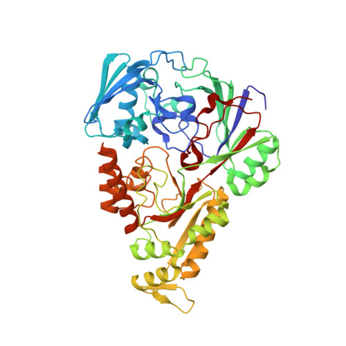
    In human pathogenic bacteria, nickel is required for the activation of two enzymes, urease and [NiFe]-hydrogenase, necessary for host infection. Acquisition of Ni(II) is mediated by either permeases or ABC-importers, the latter including a subclass that involves an extracytoplasmic nickel-binding protein, Ni-BP. This study reports on the structure of three Ni-BPs from a diversity of human pathogens and on the existence of three new nickel-binding motifs. These are different from that previously described for Escherichia coli Ni-BP NikA, known to bind nickel via a nickelophore, and indicate a variegated ligand selectivity for Ni-BPs. The structures are consistent with ligand affinities measured in solution by calorimetry and challenge the hypothesis of a general requirement of nickelophores for nickel uptake by canonical ABC importers. Phylogenetic analyses showed that Ni-BPs have different evolutionary origins and emerged independently from peptide-binding proteins, possibly explaining the promiscuous behavior of this class of Ni(II) carriers.


  • Organizational Affiliation

    University Grenoble Alpes, Institut de Biologie Structurale (IBS), 38044 Grenoble, France; CNRS, IBS, 38044 Grenoble, France; CEA, IBS, 38044 Grenoble, France.


Macromolecules
Find similar proteins by:  (by identity cutoff)  |  3D Structure
Entity ID: 1
MoleculeChains Sequence LengthOrganismDetailsImage
Putative peptide ABC-transport system periplasmic peptide-binding protein
A, B
494Campylobacter jejuni subsp. jejuni NCTC 11168 = ATCC 700819Mutation(s): 0 
Gene Names: Cj1584c
UniProt
Find proteins for Q0P844 (Campylobacter jejuni subsp. jejuni serotype O:2 (strain ATCC 700819 / NCTC 11168))
Explore Q0P844 
Go to UniProtKB:  Q0P844
Entity Groups  
Sequence Clusters30% Identity50% Identity70% Identity90% Identity95% Identity100% Identity
UniProt GroupQ0P844
Sequence Annotations
Expand
  • Reference Sequence
Small Molecules
Ligands 5 Unique
IDChains Name / Formula / InChI Key2D Diagram3D Interactions
HIS
Query on HIS

Download Ideal Coordinates CCD File 
D [auth A],
J [auth B]
HISTIDINE
C6 H10 N3 O2
HNDVDQJCIGZPNO-YFKPBYRVSA-O
PEG
Query on PEG

Download Ideal Coordinates CCD File 
F [auth A],
G [auth A],
N [auth B]
DI(HYDROXYETHYL)ETHER
C4 H10 O3
MTHSVFCYNBDYFN-UHFFFAOYSA-N
GOL
Query on GOL

Download Ideal Coordinates CCD File 
H [auth A],
L [auth B],
O [auth B]
GLYCEROL
C3 H8 O3
PEDCQBHIVMGVHV-UHFFFAOYSA-N
NI
Query on NI

Download Ideal Coordinates CCD File 
C [auth A],
I [auth B],
K [auth B]
NICKEL (II) ION
Ni
VEQPNABPJHWNSG-UHFFFAOYSA-N
MG
Query on MG

Download Ideal Coordinates CCD File 
E [auth A],
M [auth B]
MAGNESIUM ION
Mg
JLVVSXFLKOJNIY-UHFFFAOYSA-N
Experimental Data & Validation

Experimental Data

  • Method: X-RAY DIFFRACTION
  • Resolution: 2.20 Å
  • R-Value Free: 0.241 
  • R-Value Work: 0.192 
  • R-Value Observed: 0.195 
  • Space Group: C 1 2 1
Unit Cell:
Length ( Å )Angle ( ˚ )
a = 154.48α = 90
b = 72.77β = 111.99
c = 86.09γ = 90
Software Package:
Software NamePurpose
PHASERphasing
PHENIXrefinement
XDSdata reduction
XSCALEdata scaling

Structure Validation

View Full Validation Report


Ligand Structure Quality Assessment 


Entry History 

Deposition Data

Revision History  (Full details and data files)

  • Version 1.0: 2014-10-01
    Type: Initial release
  • Version 1.1: 2014-11-19
    Changes: Database references
  • Version 1.2: 2023-09-20
    Changes: Data collection, Database references, Derived calculations, Refinement description