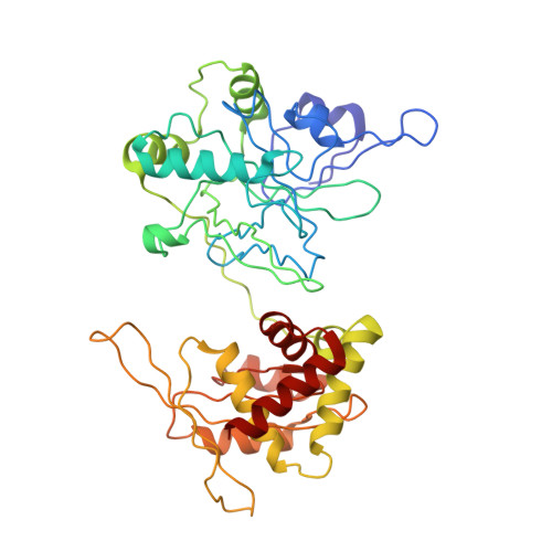
Structure and mutagenesis of the DNA modification-dependent restriction endonuclease AspBHI.
Horton, J.R., Nugent, R.L., Li, A., Mabuchi, M.Y., Fomenkov, A., Cohen-Karni, D., Griggs, R.M., Zhang, X., Wilson, G.G., Zheng, Y., Xu, S.Y., Cheng, X.(2014) Sci Rep 4: 4246-4246
- PubMed: 24604015
- DOI: https://doi.org/10.1038/srep04246
- Primary Citation of Related Structures:
4OC8 - PubMed Abstract:
The modification-dependent restriction endonuclease AspBHI recognizes 5-methylcytosine (5mC) in the double-strand DNA sequence context of (C/T)(C/G)(5mC)N(C/G) (N = any nucleotide) and cleaves the two strands a fixed distance (N12/N16) 3' to the modified cytosine. We determined the crystal structure of the homo-tetrameric AspBHI. Each subunit of the protein comprises two domains: an N-terminal DNA-recognition domain and a C-terminal DNA cleavage domain. The N-terminal domain is structurally similar to the eukaryotic SET and RING-associated (SRA) domain, which is known to bind to a hemi-methylated CpG dinucleotide. The C-terminal domain is structurally similar to classic Type II restriction enzymes and contains the endonuclease catalytic-site motif of DX20EAK. To understand how specific amino acids affect AspBHI recognition preference, we generated a homology model of the AspBHI-DNA complex, and probed the importance of individual amino acids by mutagenesis. Ser41 and Arg42 are predicted to be located in the DNA minor groove 5' to the modified cytosine. Substitution of Ser41 with alanine (S41A) and cysteine (S41C) resulted in mutants with altered cleavage activity. All 19 Arg42 variants resulted in loss of endonuclease activity.
Organizational Affiliation:
Department of Biochemistry, Emory University School of Medicine, 1510 Clifton Road, Atlanta, Georgia 30322, USA.