4O9M

Human DNA polymerase beta complexed with adenylated tetrahydrofuran (abasic site) containing DNA


Experimental Data Snapshot

  • Method: X-RAY DIFFRACTION
  • Resolution: 2.29 Å
  • R-Value Free: 0.261 
  • R-Value Work: 0.193 
  • R-Value Observed: 0.200 

wwPDB Validation   3D Report Full Report


Ligand Structure Quality Assessment 


This is version 1.2 of the entry. See complete history


Literature

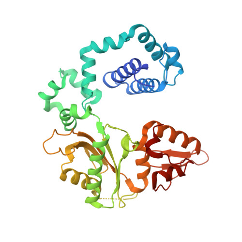
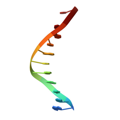
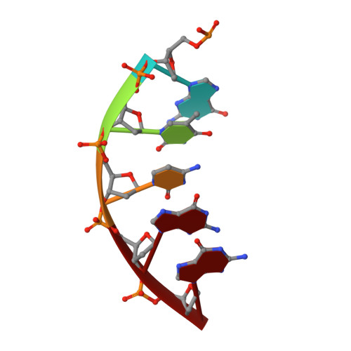
Role of polymerase beta in complementing aprataxin deficiency during abasic-site base excision repair.

Caglayan, M.Batra, V.K.Sassa, A.Prasad, R.Wilson, S.H.

(2014) Nat Struct Mol Biol 21: 497-499

  • DOI: https://doi.org/10.1038/nsmb.2818
  • Primary Citation of Related Structures:  
    4O9M

  • PubMed Abstract: 

    DNA polymerase β (pol β) lyase removal of 5'-deoxyribose phosphate (5'-dRP) from base excision repair (BER) intermediates is critical in mammalian BER involving the abasic site. We found that pol β also removes 5'-adenylated dRP from BER intermediates after abortive ligation. The crystal structure of a human pol β-DNA complex showed the 5'-AMP-dRP group positioned in the lyase active site. Pol β expression rescued methyl methanesulfonate sensitivity in aprataxin (hnt3)- and FEN1 (rad27)-deficient yeast.


  • Organizational Affiliation

    Laboratory of Structural Biology, National Institute of Environmental Health Sciences, National Institutes of Health, Research Triangle Park, North Carolina, USA.


Macromolecules

Find similar proteins by:  (by identity cutoff)  |  3D Structure
Entity ID: 4
MoleculeChains Sequence LengthOrganismDetailsImage
DNA polymerase betaD [auth A]335Homo sapiensMutation(s): 0 
Gene Names: POLB
EC: 2.7.7.7 (PDB Primary Data), 4.2.99 (PDB Primary Data)
UniProt & NIH Common Fund Data Resources
Find proteins for P06746 (Homo sapiens)
Explore P06746 
Go to UniProtKB:  P06746
PHAROS:  P06746
GTEx:  ENSG00000070501 
Entity Groups  
Sequence Clusters30% Identity50% Identity70% Identity90% Identity95% Identity100% Identity
UniProt GroupP06746
Sequence Annotations
Expand
  • Reference Sequence

Find similar nucleic acids by:  Sequence   |   3D Structure  

Entity ID: 1
MoleculeChains LengthOrganismImage
DNA (5'-D(*CP*CP*GP*AP*CP*AP*GP*CP*GP*CP*AP*TP*CP*AP*GP*C)-3')A [auth B]16N/A
Sequence Annotations
Expand
  • Reference Sequence

Find similar nucleic acids by:  Sequence   |   3D Structure  

Entity ID: 2
MoleculeChains LengthOrganismImage
DNA (5'-D(*GP*CP*TP*GP*AP*TP*GP*CP*GP*CP*C)-3')B [auth C]11N/A
Sequence Annotations
Expand
  • Reference Sequence

Find similar nucleic acids by:  Sequence   |   3D Structure  

Entity ID: 3
MoleculeChains LengthOrganismImage
(5'-D(P*GP*TP*CP*GP*G)-3');C [auth D]5N/A
Sequence Annotations
Expand
  • Reference Sequence
Small Molecules
Ligands 2 Unique
IDChains Name / Formula / InChI Key2D Diagram3D Interactions
2RW
Query on 2RW

Download Ideal Coordinates CCD File 
E [auth D][(2R,3S,4R,5R)-5-(6-amino-9H-purin-9-yl)-3,4-dihydroxytetrahydrofuran-2-yl]methyl [(2R,3S)-3-hydroxytetrahydrofuran-2-yl]methyl dihydrogen diphosphate
C15 H23 N5 O12 P2
ZJGNPGFGXQKXSY-IBCGMDAMSA-N
NA
Query on NA

Download Ideal Coordinates CCD File 
F [auth A],
G [auth A]
SODIUM ION
Na
FKNQFGJONOIPTF-UHFFFAOYSA-N
Experimental Data & Validation

Experimental Data

  • Method: X-RAY DIFFRACTION
  • Resolution: 2.29 Å
  • R-Value Free: 0.261 
  • R-Value Work: 0.193 
  • R-Value Observed: 0.200 
  • Space Group: P 1 21 1
Unit Cell:
Length ( Å )Angle ( ˚ )
a = 54.627α = 90
b = 78.873β = 105.94
c = 54.324γ = 90
Software Package:
Software NamePurpose
HKL-2000data collection
PHENIXmodel building
PHENIXrefinement
HKL-2000data reduction
HKL-2000data scaling
PHENIXphasing

Structure Validation

View Full Validation Report


Ligand Structure Quality Assessment 


Entry History 

Deposition Data

Revision History  (Full details and data files)

  • Version 1.0: 2014-04-30
    Type: Initial release
  • Version 1.1: 2014-06-11
    Changes: Database references
  • Version 1.2: 2023-09-20
    Changes: Data collection, Database references, Derived calculations, Refinement description