


X-ray Crystal Structure of Isovaleryl-CoA Dehydrogenase from Brucella suis
Fairman, J.W., Edwards, T.E., Lorimer, D.To be published.
Experimental Data Snapshot
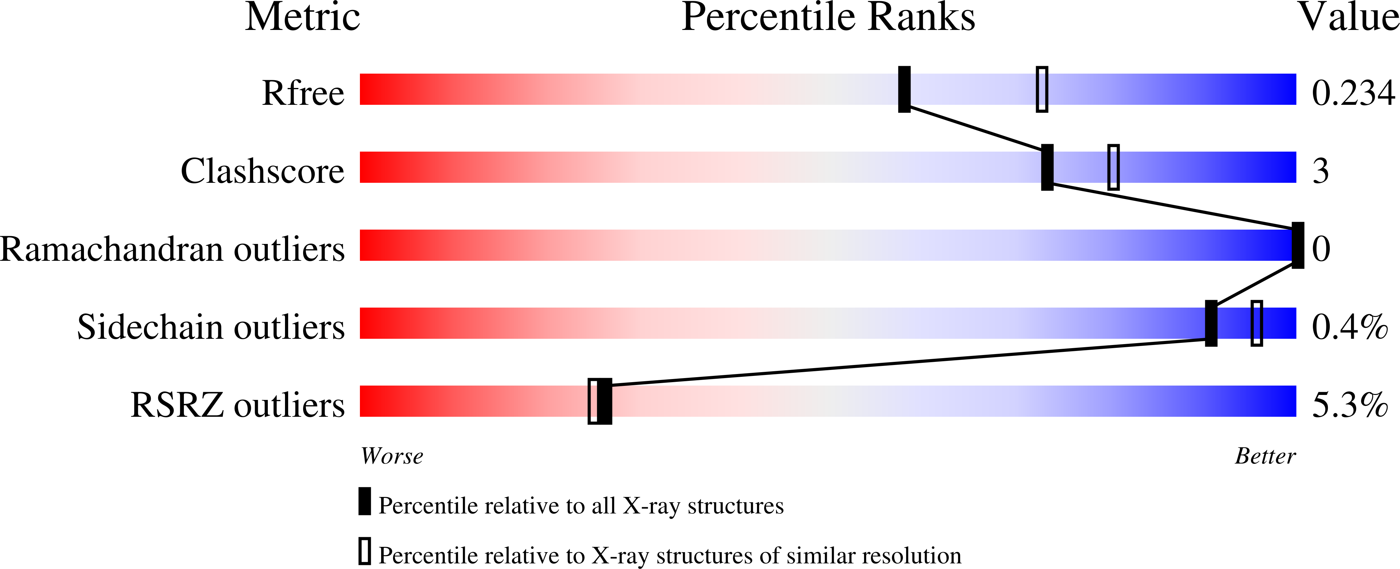
wwPDB Validation 3D Report Full Report
Entity ID: 1 | |||||
|---|---|---|---|---|---|
| Molecule | Chains | Sequence Length | Organism | Details | Image |
| Isovaleryl-CoA dehydrogenase | 390 | Brucella suis 1330 | Mutation(s): 0 Gene Names: ivd, BR0020, BS1330_I0020 EC: 1.3.99.10 | 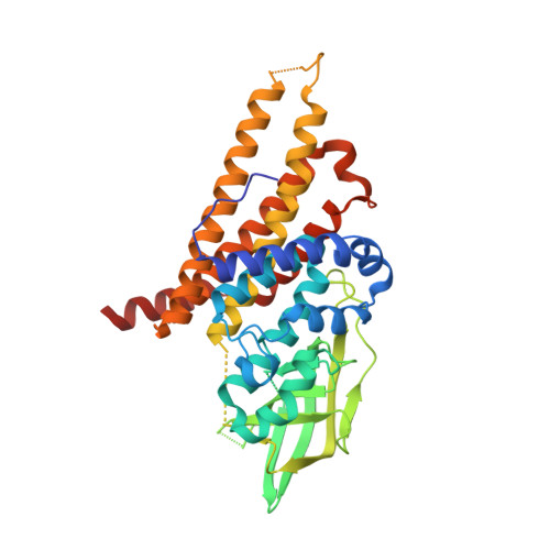 | |
UniProt | |||||
Find proteins for A0A0H3G544 (Brucella suis biovar 1 (strain 1330)) Explore A0A0H3G544 Go to UniProtKB: A0A0H3G544 | |||||
Entity Groups | |||||
| Sequence Clusters | 30% Identity50% Identity70% Identity90% Identity95% Identity100% Identity | ||||
| UniProt Group | A0A0H3G544 | ||||
Sequence AnnotationsExpand | |||||
| |||||
| Ligands 2 Unique | |||||
|---|---|---|---|---|---|
| ID | Chains | Name / Formula / InChI Key | 2D Diagram | 3D Interactions | |
| PG5 Query on PG5 | F [auth B], G [auth C], H [auth D] | 1-METHOXY-2-[2-(2-METHOXY-ETHOXY]-ETHANE C8 H18 O4 YFNKIDBQEZZDLK-UHFFFAOYSA-N |  | ||
| CA Query on CA | E [auth A] | CALCIUM ION Ca BHPQYMZQTOCNFJ-UHFFFAOYSA-N |  | ||
| Length ( Å ) | Angle ( ˚ ) |
|---|---|
| a = 82.21 | α = 90 |
| b = 91.07 | β = 99.25 |
| c = 102.14 | γ = 90 |
| Software Name | Purpose |
|---|---|
| XSCALE | data scaling |
| PHASER | phasing |
| PHENIX | refinement |
| PDB_EXTRACT | data extraction |
| XDS | data reduction |
RCSB PDB (citation) is hosted by
RCSB PDB is a member of the

