4O3M

Ternary complex of Bloom's syndrome helicase


Experimental Data Snapshot

  • Method: X-RAY DIFFRACTION
  • Resolution: 2.30 Å
  • R-Value Free: 0.215 
  • R-Value Work: 0.181 
  • R-Value Observed: 0.183 

wwPDB Validation   3D Report Full Report


Ligand Structure Quality Assessment 


This is version 1.3 of the entry. See complete history


Literature

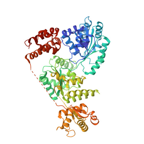
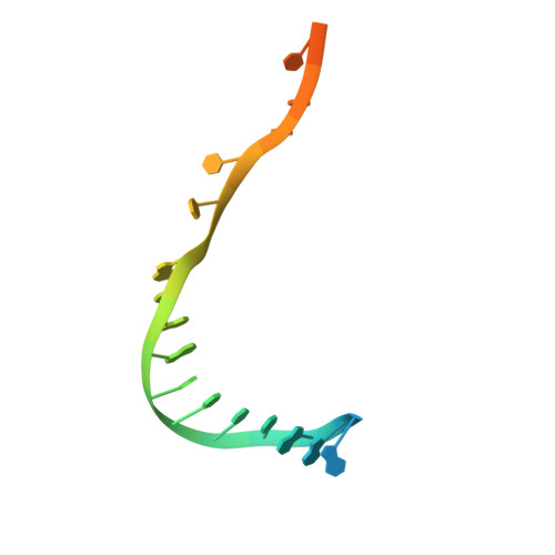
Structure of human Bloom's syndrome helicase in complex with ADP and duplex DNA.

Swan, M.K.Legris, V.Tanner, A.Reaper, P.M.Vial, S.Bordas, R.Pollard, J.R.Charlton, P.A.Golec, J.M.Bertrand, J.A.

(2014) Acta Crystallogr D Biol Crystallogr 70: 1465-1475

  • DOI: https://doi.org/10.1107/S139900471400501X
  • Primary Citation of Related Structures:  
    4O3M

  • PubMed Abstract: 

    Bloom's syndrome is an autosomal recessive genome-instability disorder associated with a predisposition to cancer, premature aging and developmental abnormalities. It is caused by mutations that inactivate the DNA helicase activity of the BLM protein or nullify protein expression. The BLM helicase has been implicated in the alternative lengthening of telomeres (ALT) pathway, which is essential for the limitless replication of some cancer cells. This pathway is used by 10-15% of cancers, where inhibitors of BLM are expected to facilitate telomere shortening, leading to apoptosis or senescence. Here, the crystal structure of the human BLM helicase in complex with ADP and a 3'-overhang DNA duplex is reported. In addition to the helicase core, the BLM construct used for crystallization (residues 640-1298) includes the RecQ C-terminal (RQC) and the helicase and ribonuclease D C-terminal (HRDC) domains. Analysis of the structure provides detailed information on the interactions of the protein with DNA and helps to explain the mechanism coupling ATP hydrolysis and DNA unwinding. In addition, mapping of the missense mutations onto the structure provides insights into the molecular basis of Bloom's syndrome.


  • Organizational Affiliation

    Vertex Pharmaceuticals (Europe), Abingdon, Oxfordshire, England.


Macromolecules

Find similar proteins by:  (by identity cutoff)  |  3D Structure
Entity ID: 1
MoleculeChains Sequence LengthOrganismDetailsImage
Bloom syndrome protein659Homo sapiensMutation(s): 0 
Gene Names: BLMRECQ2RECQL3
EC: 3.6.4.12
UniProt & NIH Common Fund Data Resources
Find proteins for P54132 (Homo sapiens)
Explore P54132 
Go to UniProtKB:  P54132
PHAROS:  P54132
GTEx:  ENSG00000197299 
Entity Groups  
Sequence Clusters30% Identity50% Identity70% Identity90% Identity95% Identity100% Identity
UniProt GroupP54132
Sequence Annotations
Expand
  • Reference Sequence

Find similar nucleic acids by:  Sequence   |   3D Structure  

Entity ID: 2
MoleculeChains LengthOrganismImage
5'-D(*AP*GP*CP*GP*TP*CP*GP*AP*GP*AP*TP*CP*CP*AP*AP*G)-3'B [auth P]16N/A
Sequence Annotations
Expand
  • Reference Sequence

Find similar nucleic acids by:  Sequence   |   3D Structure  

Entity ID: 3
MoleculeChains LengthOrganismImage
5'-D(*CP*TP*TP*GP*GP*AP*TP*CP*TP*CP*GP*AP*CP*GP*CP*TP*CP*TP*CP*CP*CP*TP*TP*A)-3'C [auth T]24N/A
Sequence Annotations
Expand
  • Reference Sequence
Small Molecules
Ligands 4 Unique
IDChains Name / Formula / InChI Key2D Diagram3D Interactions
ADP
Query on ADP

Download Ideal Coordinates CCD File 
F [auth A]ADENOSINE-5'-DIPHOSPHATE
C10 H15 N5 O10 P2
XTWYTFMLZFPYCI-KQYNXXCUSA-N
ZN
Query on ZN

Download Ideal Coordinates CCD File 
E [auth A]ZINC ION
Zn
PTFCDOFLOPIGGS-UHFFFAOYSA-N
EDO
Query on EDO

Download Ideal Coordinates CCD File 
G [auth A]
H [auth A]
I [auth A]
J [auth A]
K [auth A]
G [auth A],
H [auth A],
I [auth A],
J [auth A],
K [auth A],
L [auth A],
M [auth A],
N [auth A]
1,2-ETHANEDIOL
C2 H6 O2
LYCAIKOWRPUZTN-UHFFFAOYSA-N
CA
Query on CA

Download Ideal Coordinates CCD File 
D [auth A]CALCIUM ION
Ca
BHPQYMZQTOCNFJ-UHFFFAOYSA-N
Experimental Data & Validation

Experimental Data

  • Method: X-RAY DIFFRACTION
  • Resolution: 2.30 Å
  • R-Value Free: 0.215 
  • R-Value Work: 0.181 
  • R-Value Observed: 0.183 
  • Space Group: C 1 2 1
Unit Cell:
Length ( Å )Angle ( ˚ )
a = 100.93α = 90
b = 164.68β = 90.14
c = 50.98γ = 90
Software Package:
Software NamePurpose
SHARPphasing
BUSTERrefinement
XDSdata reduction
XDSdata scaling

Structure Validation

View Full Validation Report


Ligand Structure Quality Assessment 


Entry History 

Deposition Data

Revision History  (Full details and data files)

  • Version 1.0: 2014-03-12
    Type: Initial release
  • Version 1.1: 2014-07-02
    Changes: Database references
  • Version 1.2: 2017-11-22
    Changes: Refinement description
  • Version 1.3: 2023-09-20
    Changes: Data collection, Database references, Derived calculations, Refinement description