4O3B

Crystal structure of an open/closed glua2 ligand-binding domain dimer at 1.91 A resolution


Experimental Data Snapshot

  • Method: X-RAY DIFFRACTION
  • Resolution: 1.91 Å
  • R-Value Free: 0.178 
  • R-Value Work: 0.138 
  • R-Value Observed: 0.140 

wwPDB Validation   3D Report Full Report


This is version 1.4 of the entry. See complete history


Literature

L-Asp is a useful tool in the purification of the ionotropic glutamate receptor A2 ligand-binding domain.

Krintel, C.Frydenvang, K.Ceravalls de Rabassa, A.Kaern, A.M.Gajhede, M.Pickering, D.S.Kastrup, J.S.

(2014) FEBS J 281: 2422-2430

  • DOI: https://doi.org/10.1111/febs.12795
  • Primary Citation of Related Structures:  
    4O3A, 4O3B, 4O3C

  • PubMed Abstract: 

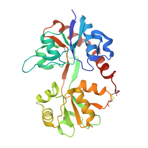
    In purification of the ionotropic glutamate receptor A2 (GluA2) ligand-binding domain (LBD), L-Glu-supplemented buffers have previously been used for protein stabilization during the procedure. This sometimes hampers structural studies of low-affinity ligands, because L-Glu is difficult to displace, despite extensive dialysis. Here, we show that L-Asp binds to full-length GluA2 with low affinity (Ki = 0.63 mM) and to the GluA2 LBD with even lower affinity (Ki = 2.6 mM), and we use differential scanning fluorimetry to show that L-Asp is able to stabilize the isolated GluA2 LBD. We also show that L-Asp can replace L-Glu during purification, providing both equal yields and purity of the resulting protein sample. Furthermore, we solved three structures of the GluA2 LBD in the presence of 7.5, 50 and 250 mM L-Asp. Surprisingly, with 7.5 mM L-Asp, the GluA2 LBD crystallized as a mixed dimer, with L-Glu being present in one subunit, and neither L-Asp nor L-Glu being present in the other subunit. Thus, residual L-Glu is retained from the expression medium. On the other hand, only L-Asp was found at the binding site when 50 or 250 mM L-Asp was used for crystallization. The binding mode observed for L-Asp at the GluA2 LBD is very similar to that described for L-Glu. Taking our findings together, we have shown that L-Asp can be used instead of L-Glu for ligand-dependent stabilization of the GluA2 LBD during purification. This will enable structural studies of low-affinity ligands for lead optimization in structure-based drug design.


  • Organizational Affiliation

    Department of Drug Design and Pharmacology, Faculty of Health and Medical Sciences, University of Copenhagen, Denmark.


Macromolecules
Find similar proteins by:  (by identity cutoff)  |  3D Structure
Entity ID: 1
MoleculeChains Sequence LengthOrganismDetailsImage
Glutamate receptor 2
A, B
263Rattus norvegicusMutation(s): 0 
Gene Names: Gria2Glur2
UniProt
Find proteins for P19491 (Rattus norvegicus)
Explore P19491 
Go to UniProtKB:  P19491
Entity Groups  
Sequence Clusters30% Identity50% Identity70% Identity90% Identity95% Identity100% Identity
UniProt GroupP19491
Sequence Annotations
Expand
  • Reference Sequence
Small Molecules
Ligands 7 Unique
IDChains Name / Formula / InChI Key2D Diagram3D Interactions
PG4
Query on PG4

Download Ideal Coordinates CCD File 
Q [auth B]TETRAETHYLENE GLYCOL
C8 H18 O5
UWHCKJMYHZGTIT-UHFFFAOYSA-N
GLU
Query on GLU

Download Ideal Coordinates CCD File 
L [auth B]GLUTAMIC ACID
C5 H9 N O4
WHUUTDBJXJRKMK-VKHMYHEASA-N
PEG
Query on PEG

Download Ideal Coordinates CCD File 
J [auth A]DI(HYDROXYETHYL)ETHER
C4 H10 O3
MTHSVFCYNBDYFN-UHFFFAOYSA-N
SO4
Query on SO4

Download Ideal Coordinates CCD File 
C [auth A],
D [auth A],
E [auth A],
M [auth B]
SULFATE ION
O4 S
QAOWNCQODCNURD-UHFFFAOYSA-L
GOL
Query on GOL

Download Ideal Coordinates CCD File 
F [auth A]
G [auth A]
H [auth A]
N [auth B]
O [auth B]
F [auth A],
G [auth A],
H [auth A],
N [auth B],
O [auth B],
P [auth B]
GLYCEROL
C3 H8 O3
PEDCQBHIVMGVHV-UHFFFAOYSA-N
ACT
Query on ACT

Download Ideal Coordinates CCD File 
K [auth A]ACETATE ION
C2 H3 O2
QTBSBXVTEAMEQO-UHFFFAOYSA-M
CL
Query on CL

Download Ideal Coordinates CCD File 
I [auth A]CHLORIDE ION
Cl
VEXZGXHMUGYJMC-UHFFFAOYSA-M
Binding Affinity Annotations 
IDSourceBinding Affinity
GLU Binding MOAD:  4O3B Ki: 820 (nM) from 1 assay(s)
Experimental Data & Validation

Experimental Data

  • Method: X-RAY DIFFRACTION
  • Resolution: 1.91 Å
  • R-Value Free: 0.178 
  • R-Value Work: 0.138 
  • R-Value Observed: 0.140 
  • Space Group: P 21 21 21
Unit Cell:
Length ( Å )Angle ( ˚ )
a = 62.72α = 90
b = 93.72β = 90
c = 97.14γ = 90
Software Package:
Software NamePurpose
PHASERphasing
PHENIXrefinement
PDB_EXTRACTdata extraction
MOSFLMdata reduction
SCALAdata scaling

Structure Validation

View Full Validation Report


Entry History 

Deposition Data

Revision History  (Full details and data files)

  • Version 1.0: 2014-04-16
    Type: Initial release
  • Version 1.1: 2014-06-18
    Changes: Database references
  • Version 1.2: 2017-05-31
    Changes: Structure summary
  • Version 1.3: 2018-03-07
    Changes: Data collection
  • Version 1.4: 2023-09-20
    Changes: Data collection, Database references, Derived calculations, Refinement description