4NYC

Crystal structure of the E. coli thiM riboswitch in complex with thieno[2,3-b]pyrazin-7-amine


Experimental Data Snapshot

  • Method: X-RAY DIFFRACTION
  • Resolution: 3.15 Å
  • R-Value Free: 0.264 
  • R-Value Work: 0.202 

wwPDB Validation   3D Report Full Report


Ligand Structure Quality Assessment 


This is version 1.1 of the entry. See complete history


Literature

Validating Fragment-Based Drug Discovery for Biological RNAs: Lead Fragments Bind and Remodel the TPP Riboswitch Specifically.

Warner, K.D.Homan, P.Weeks, K.M.Smith, A.G.Abell, C.Ferre-D'Amare, A.R.

(2014) Chem Biol 21: 591-595

  • DOI: https://doi.org/10.1016/j.chembiol.2014.03.007
  • Primary Citation of Related Structures:  
    4NYA, 4NYB, 4NYC, 4NYD, 4NYG

  • PubMed Abstract: 

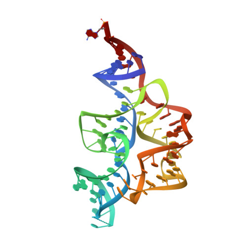
    Thiamine pyrophosphate (TPP) riboswitches regulate essential genes in bacteria by changing conformation upon binding intracellular TPP. Previous studies using fragment-based approaches identified small molecule "fragments" that bind this gene-regulatory mRNA domain. Crystallographic studies now show that, despite having micromolar Kds, four different fragments bind the TPP riboswitch site-specifically, occupying the pocket that recognizes the aminopyrimidine of TPP. Unexpectedly, the unoccupied site that would recognize the pyrophosphate of TPP rearranges into a structure distinct from that of the cognate complex. This idiosyncratic fragment-induced conformation, also characterized by small-angle X-ray scattering and chemical probing, represents a possible mechanism for adventitious ligand discrimination by the riboswitch, and suggests that off-pathway conformations of RNAs can be targeted for drug development. Our structures, together with previous screening studies, demonstrate the feasibility of fragment-based drug discovery against RNA targets.


  • Organizational Affiliation

    National Heart, Lung and Blood Institute, 50 South Drive, MSC 8012, Bethesda, MD 20892-8012, USA; Department of Chemistry, University of Cambridge, Lensfield Road, Cambridge CB2 1EW, UK.


Macromolecules
Find similar nucleic acids by:  (by identity cutoff)  |  3D Structure
Entity ID: 1
MoleculeChains LengthOrganismImage
thiM TPP riboswitch83Escherichia coli
Sequence Annotations
Expand
  • Reference Sequence
Small Molecules
Ligands 3 Unique
IDChains Name / Formula / InChI Key2D Diagram3D Interactions
SVN
Query on SVN

Download Ideal Coordinates CCD File 
B [auth A]thieno[2,3-b]pyrazin-7-amine
C6 H5 N3 S
ZRVKSPNBHZCQKY-UHFFFAOYSA-N
MN
Query on MN

Download Ideal Coordinates CCD File 
D [auth A],
E [auth A],
H [auth A],
J [auth A],
K [auth A]
MANGANESE (II) ION
Mn
WAEMQWOKJMHJLA-UHFFFAOYSA-N
MG
Query on MG

Download Ideal Coordinates CCD File 
C [auth A]
F [auth A]
G [auth A]
I [auth A]
L [auth A]
C [auth A],
F [auth A],
G [auth A],
I [auth A],
L [auth A],
M [auth A],
N [auth A],
O [auth A],
P [auth A],
Q [auth A],
R [auth A]
MAGNESIUM ION
Mg
JLVVSXFLKOJNIY-UHFFFAOYSA-N
Experimental Data & Validation

Experimental Data

  • Method: X-RAY DIFFRACTION
  • Resolution: 3.15 Å
  • R-Value Free: 0.264 
  • R-Value Work: 0.202 
  • Space Group: P 32 1 2
Unit Cell:
Length ( Å )Angle ( ˚ )
a = 65.122α = 90
b = 65.122β = 90
c = 101.975γ = 120
Software Package:
Software NamePurpose
SCALEPACKdata scaling
CNSrefinement
PDB_EXTRACTdata extraction
HKL-2000data collection
DENZOdata reduction
PHASERphasing

Structure Validation

View Full Validation Report


Ligand Structure Quality Assessment 


Entry History 

Deposition Data

Revision History  (Full details and data files)

  • Version 1.0: 2014-06-04
    Type: Initial release
  • Version 1.1: 2023-09-20
    Changes: Data collection, Database references, Derived calculations, Refinement description