4NY4

Crystal structure of CYP3A4 in complex with an inhibitor


Experimental Data Snapshot

  • Method: X-RAY DIFFRACTION
  • Resolution: 2.95 Å
  • R-Value Free: 0.273 
  • R-Value Work: 0.249 
  • R-Value Observed: 0.250 

wwPDB Validation   3D Report Full Report


Ligand Structure Quality Assessment 


This is version 1.1 of the entry. See complete history


Literature

Structure-based ligand design to overcome CYP inhibition in drug discovery projects.

Branden, G.Sjogren, T.Schnecke, V.Xue, Y.

(2014) Drug Discov Today 19: 905-911

  • DOI: https://doi.org/10.1016/j.drudis.2014.03.012
  • Primary Citation of Related Structures:  
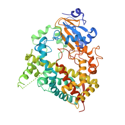
    4NY4, 4NZ2

  • PubMed Abstract: 

    Cytochrome P450 (CYP) enzymes are key players in xenobiotic metabolism, and inhibition of CYPs can therefore result in unwanted drug-drug interactions. Within drug discovery, CYP inhibition can cause delays in the progression of candidate drugs, or even premature closure of projects. During the past decade, a massive effort in the pharmaceutical industry and academic research has produced a wealth of structural information in the CYP field. In this short review, we will describe how structure-based approaches can be used in the pharmaceutical industry to work away from CYP inhibition, with a focus on the opportunities and challenges. We will show two examples from our own work where structural information on CYP2C9 and CYP3A4 inhibitor complexes have been successfully exploited in ongoing drug discovery projects.


  • Organizational Affiliation

    Department of Chemistry and Molecular Biology, University of Gothenburg, Göteborg S-405 30, Sweden. Electronic address: gisela.branden@gu.se.


Macromolecules
Find similar proteins by:  (by identity cutoff)  |  3D Structure
Entity ID: 1
MoleculeChains Sequence LengthOrganismDetailsImage
Cytochrome P450 3A4484Homo sapiensMutation(s): 0 
Gene Names: CYP3A3CYP3A4CYP3A4 CYP3A3
EC: 1.14.13 (PDB Primary Data), 1.14.13.157 (PDB Primary Data), 1.14.13.32 (PDB Primary Data), 1.14.13.67 (PDB Primary Data), 1.14.13.97 (PDB Primary Data)
UniProt & NIH Common Fund Data Resources
Find proteins for P08684 (Homo sapiens)
Explore P08684 
Go to UniProtKB:  P08684
PHAROS:  P08684
GTEx:  ENSG00000160868 
Entity Groups  
Sequence Clusters30% Identity50% Identity70% Identity90% Identity95% Identity100% Identity
UniProt GroupP08684
Sequence Annotations
Expand
  • Reference Sequence
Small Molecules
Ligands 2 Unique
IDChains Name / Formula / InChI Key2D Diagram3D Interactions
HEM
Query on HEM

Download Ideal Coordinates CCD File 
B [auth A]PROTOPORPHYRIN IX CONTAINING FE
C34 H32 Fe N4 O4
KABFMIBPWCXCRK-RGGAHWMASA-L
2QH
Query on 2QH

Download Ideal Coordinates CCD File 
C [auth A](8R)-3,3-difluoro-8-[4-fluoro-3-(pyridin-3-yl)phenyl]-8-(4-methoxy-3-methylphenyl)-2,3,4,8-tetrahydroimidazo[1,5-a]pyrimidin-6-amine
C25 H22 F3 N5 O
YGQLOGGNUHAJMB-RUZDIDTESA-N
Experimental Data & Validation

Experimental Data

  • Method: X-RAY DIFFRACTION
  • Resolution: 2.95 Å
  • R-Value Free: 0.273 
  • R-Value Work: 0.249 
  • R-Value Observed: 0.250 
  • Space Group: I 2 2 2
Unit Cell:
Length ( Å )Angle ( ˚ )
a = 78.548α = 90
b = 101.692β = 90
c = 132.422γ = 90
Software Package:
Software NamePurpose
MOSFLMdata reduction
SCALAdata scaling
PHASERphasing
BUSTER-TNTrefinement
PDB_EXTRACTdata extraction
BUSTERrefinement

Structure Validation

View Full Validation Report


Ligand Structure Quality Assessment 


Entry History 

Deposition Data

Revision History  (Full details and data files)

  • Version 1.0: 2014-08-13
    Type: Initial release
  • Version 1.1: 2024-02-28
    Changes: Data collection, Database references, Derived calculations