4NXS

Crystal structure of human alpha-galactosidase A in complex with 1-deoxygalactonojirimycin-pFPhT


Experimental Data Snapshot

  • Method: X-RAY DIFFRACTION
  • Resolution: 2.55 Å
  • R-Value Free: 0.232 
  • R-Value Work: 0.187 
  • R-Value Observed: 0.190 

wwPDB Validation   3D Report Full Report


Ligand Structure Quality Assessment 


This is version 2.1 of the entry. See complete history


Literature

Molecular Basis of 1-Deoxygalactonojirimycin Arylthiourea Binding to Human alpha-Galactosidase A: Pharmacological Chaperoning Efficacy on Fabry Disease Mutants.

Yu, Y.Mena-Barragan, T.Higaki, K.Johnson, J.L.Drury, J.E.Lieberman, R.L.Nakasone, N.Ninomiya, H.Tsukimura, T.Sakuraba, H.Suzuki, Y.Nanba, E.Mellet, C.O.Garcia Fernandez, J.M.Ohno, K.

(2014) ACS Chem Biol 9: 1460-1469

  • DOI: https://doi.org/10.1021/cb500143h
  • Primary Citation of Related Structures:  
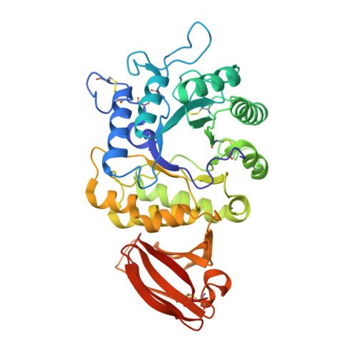
    4NXS

  • PubMed Abstract: 

    Fabry disease (FD) is an X-linked lysosomal storage disorder caused by mutations in the GLA gene often leading to missense α-galactosidase A (α-Gal A) variants that undergo premature endoplasmic reticulum-associated degradation due to folding defects. We have synthesized and characterized a new family of neutral amphiphilic pharmacological chaperones, namely 1-deoxygalactonojirimycin-arylthioureas (DGJ-ArTs), capable of stabilizing α-Gal A and restoring trafficking. Binding to the enzyme is reinforced by a strong hydrogen bond involving the aryl-N'H thiourea proton and the catalytic aspartic acid acid D231 of α-Gal A, as confirmed by a 2.55 Å resolution cocrystal structure. Selected candidates enhanced α-Gal A activity and ameliorate globotriaosylceramide (Gb3) accumulation and autophagy impairments in FD cell cultures. Moreover, they acted synergistically with the proteostasis regulator 4-phenylbutyric acid, appearing to be promising leads as pharmacological chaperones for FD.


  • Organizational Affiliation

    Division of Functional Genomics, Research Center for Bioscience and Technology, Tottori University , Yonago 683-8503, Japan.


Macromolecules
Find similar proteins by:  (by identity cutoff)  |  3D Structure
Entity ID: 1
MoleculeChains Sequence LengthOrganismDetailsImage
Alpha-galactosidase A
A, B
398Homo sapiensMutation(s): 0 
Gene Names: GLA
EC: 3.2.1.22
UniProt & NIH Common Fund Data Resources
Find proteins for P06280 (Homo sapiens)
Explore P06280 
Go to UniProtKB:  P06280
PHAROS:  P06280
GTEx:  ENSG00000102393 
Entity Groups  
Sequence Clusters30% Identity50% Identity70% Identity90% Identity95% Identity100% Identity
UniProt GroupP06280
Sequence Annotations
Expand
  • Reference Sequence
Oligosaccharides

Help

Entity ID: 2
MoleculeChains Length2D Diagram Glycosylation3D Interactions
2-acetamido-2-deoxy-beta-D-glucopyranose-(1-4)-2-acetamido-2-deoxy-beta-D-glucopyranose
C, D
2N-Glycosylation
Glycosylation Resources
GlyTouCan:  G42666HT
GlyCosmos:  G42666HT
GlyGen:  G42666HT
Small Molecules
Ligands 3 Unique
IDChains Name / Formula / InChI Key2D Diagram3D Interactions
2OZ
Query on 2OZ

Download Ideal Coordinates CCD File 
J [auth A],
O [auth B]
(2R,3S,4R,5S)-N-(4-fluorophenyl)-3,4,5-trihydroxy-2-(hydroxymethyl)piperidine-1-carbothioamide
C13 H17 F N2 O4 S
WPBCEOLJVINCLL-NOOOWODRSA-N
NAG
Query on NAG

Download Ideal Coordinates CCD File 
H [auth A],
I [auth A],
M [auth B],
N [auth B]
2-acetamido-2-deoxy-beta-D-glucopyranose
C8 H15 N O6
OVRNDRQMDRJTHS-FMDGEEDCSA-N
SO4
Query on SO4

Download Ideal Coordinates CCD File 
E [auth A],
F [auth A],
G [auth A],
K [auth B],
L [auth B]
SULFATE ION
O4 S
QAOWNCQODCNURD-UHFFFAOYSA-L
Binding Affinity Annotations 
IDSourceBinding Affinity
2OZ BindingDB:  4NXS IC50: min: 43, max: 340 (nM) from 2 assay(s)
Experimental Data & Validation

Experimental Data

  • Method: X-RAY DIFFRACTION
  • Resolution: 2.55 Å
  • R-Value Free: 0.232 
  • R-Value Work: 0.187 
  • R-Value Observed: 0.190 
  • Space Group: P 32 2 1
Unit Cell:
Length ( Å )Angle ( ˚ )
a = 90.783α = 90
b = 90.783β = 90
c = 217.382γ = 120
Software Package:
Software NamePurpose
DENZOdata reduction
SCALEPACKdata scaling
PHASERphasing
PHENIXrefinement
PDB_EXTRACTdata extraction

Structure Validation

View Full Validation Report


Ligand Structure Quality Assessment 


Entry History 

Deposition Data

Revision History  (Full details and data files)

  • Version 1.0: 2014-06-11
    Type: Initial release
  • Version 1.1: 2014-07-30
    Changes: Database references
  • Version 1.2: 2017-11-22
    Changes: Refinement description
  • Version 2.0: 2020-07-29
    Type: Remediation
    Reason: Carbohydrate remediation
    Changes: Atomic model, Data collection, Derived calculations, Structure summary
  • Version 2.1: 2023-09-20
    Changes: Data collection, Database references, Refinement description, Structure summary