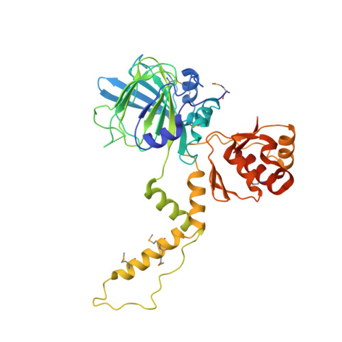
Structure and Functional Analysis of YcfD, a Novel 2-Oxoglutarate/Fe(2+)-Dependent Oxygenase Involved in Translational Regulation in Escherichia coli.
van Staalduinen, L.M., Novakowski, S.K., Jia, Z.(2014) J Mol Biol 426: 1898-1910
- PubMed: 24530688
- DOI: https://doi.org/10.1016/j.jmb.2014.02.008
- Primary Citation of Related Structures:
4NUB - PubMed Abstract:
The 2-oxoglutarate (2OG)/Fe²⁺-dependent oxygenases (2OG oxygenases) are a large family of proteins that share a similar overall three-dimensional structure and catalyze a diverse array of oxidation reactions. The Jumonji C (JmjC)-domain-containing proteins represent an important subclass of the 2OG oxygenase family that typically catalyze protein hydroxylation; however, recently, other reactions have been identified, such as tRNA modification. The Escherichia coli gene, ycfD, was predicted to be a JmjC-domain-containing protein of unknown function based on primary sequence. Recently, YcfD was determined to act as a ribosomal oxygenase, hydroxylating an arginine residue on the 50S ribosomal protein L-16 (RL-16). We have determined the crystal structure of YcfD at 2.7 Å resolution, revealing that YcfD is structurally similar to known JmjC proteins and possesses the characteristic double-stranded β-helix fold or cupin domain. Separate from the cupin domain, an additional globular module termed α-helical arm mediates dimerization of YcfD. We further have shown that 2OG binds to YcfD using isothermal titration calorimetry and identified key binding residues using mutagenesis that, together with the iron location and structural similarity with other cupin family members, allowed identification of the active site. Structural homology to ribosomal assembly proteins combined with GST (glutathione S-transferase)-YcfD pull-down of a ribosomal protein and docking of RL-16 to the YcfD active site support the role of YcfD in regulation of bacterial ribosome assembly. Furthermore, overexpression of YcfD is shown to inhibit cell growth signifying a toxic effect on ribosome assembly.
Organizational Affiliation:
Department of Biomedical and Molecular Sciences, Queen's University, Kingston, ON, K7L 3N6, Canada.